
Cytotoxicity and Limited Anti-inflammatory Responses of Licochalcone B in HaCaT Keratinocytes
Abstract
Licochalcone B is a flavonoid derived from licorice (Glycyrrhiza spp.) with reported pharmacological activities. However, its effects on skin cells remain poorly defined. Here, we examined the concentration-dependent effects of licochalcone B in human keratinocytes by assessing both its anti-inflammatory activity and cytotoxicity. In HaCaT cells stimulated with tumor necrosis factor-α and interferon-γ, low concentrations of licochalcone B modestly reduced the expression of inflammation-related chemokines, while proinflammatory cytokines were largely unchanged. In contrast, higher concentrations of licochalcone B markedly decreased cell viability and induced apoptosis, as indicated by Annexin V/propidium iodide staining, TUNEL positivity, and activation of intrinsic and extrinsic apoptotic pathways. Notably, comparable concentrations did not significantly affect the viability of hepatocyte-derived cell lines, suggesting greater susceptibility of keratinocytes to licochalcone B exposure. Collectively, these results show that licochalcone B exerts dual, concentration-dependent effects in human keratinocytes and point to a previously underexplored potential risk of skin-related toxicity associated with licorice-derived components.
Keywords:
Licochalcone B, Keratinocytes, Cytotoxicity, Chemokines, Skin toxicityIntroduction
Licorice (Glycyrrhiza spp.) is a medicinal plant extensively used in traditional medicine and functional foods, and its bioactive flavonoids have been reported to exhibit diverse pharmacological effects, including anti-inflammatory, antioxidant, hepatoprotective, neuroprotective, and anti-cancer activities.1 Nevertheless, excessive licorice consumption has been associated with adverse health effects, indicating that licorice-derived components can induce toxicity in a concentration-dependent manner.2 Importantly, studies on the toxicological properties of individual licorice-derived compounds have primarily focused on glycyrrhizin, one of the compounds from licorice, and the other components have not yet been sufficiently characterized.
Licochalcone B (LicB), a flavonoid isolated from licorice, has received considerably less attention than other licochalcones, and its biological effects are poorly understood. Several studies have suggested that LicB may exert anti-inflammatory, antioxidant, and anti-cancer activities.3–6 However, investigations focusing on its effects in skin cells are scarce. In contrast, glycyrrhizic acid and glycyrrhetic acid, which are relatively well-characterized licorice components, have been widely recognized as a major contributor to licorice-associated toxicity. As they act similarly to aldosterone, they contribute to side effects such as hypokalemia and metabolic alkalosis in licorice overdose.7–9 Another chalcone compound, licochalcone A (LicA), which is another component of licorice and shares structural similarity with LicB, has been reported to exert anti-inflammatory and antioxidant effects through pathways such as NF-κB, MAPK, JNK, and the NLRP3 inflammasome.10,11 Although LicB also belongs to the chalcone family and has been proposed to possess anti-inflammatory potential, systematic evaluations of its cytotoxicity are lacking, and to date, its effects in skin cells, particularly human keratinocytes, have not been reported.
The skin, as a primary barrier tissue directly exposed to external agents, may display distinct vulnerability to cytotoxic insults. Excessive cell death is a hallmark of cytotoxicity and contributes to tissue dysfunction and pathological conditions.12 This consideration is particularly critical in skin cells, as excessive keratinocyte death can not only compromise epidermal integrity and barrier function but also induce inflammation.13 Despite this, toxicological assessments of natural compounds in skin cells remain limited.
Therefore, the present study aimed to comprehensively characterize the biological effects of LicB in skin cells by simultaneously evaluating its potential anti-inflammatory activity and cytotoxicity. To this end, we assessed the effects of LicB in normal human keratinocytes to determine its cytotoxic profile, and in parallel, examined its anti-inflammatory potential using an atopic dermatitis-mimicking inflammatory model induced by TNF-α and IFN-γ. Given that licorice-derived chalcones are often presumed to exert beneficial anti-inflammatory effects, this dual approach allowed us to critically examine whether LicB exhibits a favorable therapeutic profile or instead poses toxic risks.
Experimental
Cell line and treatments – Human keratinocytes, namely, HaCaT cells, were incubated in a constant environment with 5% CO2 at 37°C in high-glucose Dulbecco’s Modified Eagle Medium (DMEM) (Cytiva, Marlborough, MA, USA) containing 10% fetal bovine serum (Thermo Fisher Scientific, Waltham, MA, USA) and 1% penicillin/streptomycin (Thermo Fisher Scientific, Waltham, MA, USA). The cells were subcultured every 2–3 days. LicB, which was obtained as a purified compound from Professor Jung-hyun Shim of the College of Pharmacy, Mokpo University, was dissolved in dimethyl sulfoxide (DMSO) (Sigma-Aldrich, St. Louis, MO, USA) to prepare a 10 mM stock solution. The concentration range of LicB was selected based on previous studies reporting anti-inflammatory and hepatoprotective effects within a similar range.14–16 TNF-α and IFN-γ (Bio-Techne, Minneapolis, MN, USA) were dissolved in phosphate-buffered saline (PBS; Cytiva, Marlborough, MA, USA) to prepare stock solutions of 100 and 200 μg/mL, respectively.
3-(4,5-Dimethylthiazol-2-yl)-2,5-diphenyltetrazolium bromide (MTT) assay – HaCaT cells were seeded in 96-well plates at 0.5 × 10⁴ cells/well and incubated for 16–24 h. They were treated with 2, 5, 10, and 20 μM LicB and incubated for another 24 h. They were reacted with 0.5 mg/mL MTT (Duchefa Biochemie, Haarlem, Netherlands) at 37°C in a dark environment with 5% CO2 for 40 min. After the generated formazan crystals were checked, they were dissolved in DMSO (Duchefa Biochemie, Haarlem, Netherlands). Absorbance was measured at 550 nm. Cell viability was calculated by converting the absorbance of the experimental group to a percentage compared with that of the control group.
Quantitative real-time PCR – Total RNA was extracted with 1 mL of Trizol (Thermo Fisher Scientific, Waltham, MA, USA) and purified using chloroform and isopropanol in accordance with the manufacturer’s instructions. Subsequently, 2 μg of RNA was synthesized into cDNA reacting with SuperScript IV Reverse Transcriptase (Thermo Fisher Scientific, Waltham, MA, USA). The mRNA expression of the target gene was measured with TOPrealTM qPCR 2X PreMIX (enzynomics, Daejeon, Korea), target-specific primers, and normalized to phosphoglycerate kinase 1 (PGK1), one of the housekeeping genes (Fig. S1). The primers were manufactured to have specific sequences for the mRNA of PGK1, IL-1, IL-6, IL-8, TARC, MDC, and RANTES (Table S1).
Annexin V/PI assay – The HaCaT cells exposed to 20 μM LicB for 24 or 48 h were harvested with Trypsin-EDTA (Thermo Fisher Scientific, Waltham, MA, USA) and washed with cold PBS (WELGENE, Gyeonsan, Korea) and 1X binding buffer (BD Biosciences, Franklin Lakes, NJ, USA). Then, they were stained with 5 μL of 1X Annexin V-FITC (Thermo Fisher Scientific, Waltham, MA, USA) and 5 μL of 1X propidium iodide (Thermo Fisher Scientific, Waltham, MA, USA) at room temperature for 30 and 10 min, respectively. The stained cells were suspended in 400 μL of binding buffer and analyzed via flow cytometry (Beckman Coulter, Brea, CA, USA).
TUNEL assay – The HaCaT cells treated with 20 μM LicB were subjected to a terminal deoxynucleotidyl transferase dUTP nick end labeling (TUNEL) assay (Promega, Madison, WI, USA) and detected using Cytation5 (Agilent Technologies, Santa Clara, CA, USA). In this procedure, cells were seeded in an eight-well chamber slide and stimulated with LicB for 6 h. The labeling process of the TUNEL assay was performed according to the protocol of the DeadEnd Fluorometric TUNEL System. The cells were fixed in 10% formalin (Junsei Chemical, Tokyo, Japan) for 25 min and permeabilized with 0.2% Triton X-100 (Sigma, St. Louis, MO, USA). They were rinsed with PBS and labeled with a reaction mixture containing an equilibration buffer, a nucleotide mix, and the rTdT enzyme at 37°C in a light-blocked chamber for 1 h. The labeling process was interrupted by 2X SSC. The slide was mounted with a mounting solution containing DAPI (Southern Biotech, Birmingham, AL, USA) overnight and then photographed.
Western blot analysis – The HaCaT cells were lysed using RIPA buffer (ELPIS-BIOTECH, Daejeon, Korea) containing protease inhibitors (Quartett, Berlin, Germany) and phosphatase inhibitors (Quartett, Berlin, Germany). The same amount of protein was loaded, and SDS-polyacrylamide gel electrophoresis (SDS-PAGE) was performed. The proteins in the gel were transferred to a PVDF membrane (Bio-Rad, Hercules, CA, USA), and the membrane was blocked with 5% skim milk (BD DIFCO™, Franklin Lakes, NJ, USA) in TBST with 0.05% Tween-20 (Bio-Rad, Hercules, CA, USA) at room temperature for 1 h. It was thoroughly washed and incubated with a primary antibody diluted at 1:1000 in TBST at 4°C for 16–18 h. It was washed with TBST thrice and incubated with a secondary antibody diluted at 1:3000 in TBST at room temperature for 30 min with slow shaking. The antibodies bound to the proteins were reacted with an ECL solution (Bio-Rad, Hercules, CA, USA) and detected. The primary antibodies of Bax, Bcl-2, Caspase-3, Caspase-8, and PARP were purchased from Cell Signaling TECHNOLOGY (Danvers, MA, USA); β-actin and GAPDH antibodies were procured from Santa Cruz Biotechnology (Dallas, TX, USA). The secondary antibodies were obtained from Santa Cruz Biotechnology.
Statistical analyses – All experiments were performed independently at least thrice. Data were presented as mean ± standard error (SE). Statistical significance was evaluated via t-test or one-way ANOVA, and significances between individual samples were confirmed using Tukey’s post hoc test. Data with p < 0.05 were considered statistically significant.
Results and Discussion
LicB, known for various pharmacological effects, has a chalcone structure as illustrated in Fig. 1A. To assess the impact of LicB on HaCaT cell viability, an MTT assay was performed. The survival rates of the HaCaT cells exposed to 2, 5, 10, and 20 μM LicB for 24 h decreased to 96.88%, 90.77%, 80.07%, and 43.69%, respectively (Fig. 1B). Cell viability analysis revealed a dose-dependent decrease following LicB treatment, with an estimated IC50 of approximately 18 μM in HaCaT cells. In addition, the cell morphology of 10 and 20 μM LicB treatment groups showed shrinking and degenerative changes consistent with the decline in viability (Fig. 1C). In contrast, cell viability was not markedly reduced in mouse normal hepatocytes (AML12) at the same concentration of 20 μM LicB. Notably, a slight increase of approximately 10% in cell viability was observed in human hepatoma cells (Huh7) under the same treatment conditions (Fig. S2).
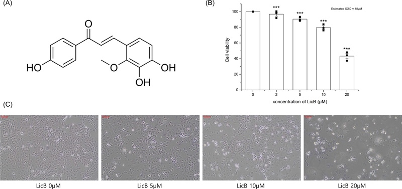
Effect of LicB on the survival of HaCaT cells. (A) Structure of LicB. (B) Cell viability of HaCaT cells exposed to different LicB concentrations (2–20 μM) for 24 h. (C) Morphological changes in HaCaT cells with 24 h exposure to different LicB concentrations (5–20 μM). The graph shows the data obtained from four independent experiments with means ± standard error (n = 4). Statistical significance was evaluated via one-way ANOVA. ***p < 0.001.
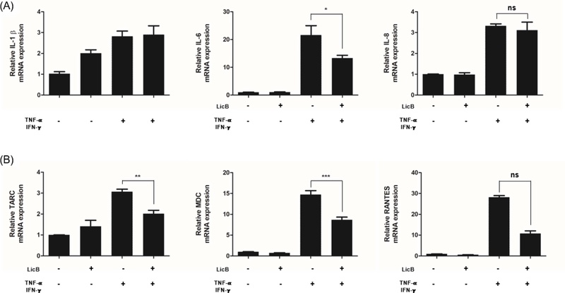
Effect of LicB on the mRNA expression of proinflammatory mediators in HaCaT cells. HaCaT cells were pretreated with 5 μM LicB, incubated for 12 h, and stimulated with 10 ng/mL TNF-α and IFN-γ. The mRNA expression levels of (A) cytokines (IL-1b, IL-6, and IL-8) and (B) chemokines (TARC, MDC, and RANTES) were measured after 24 h. Data were presented as means ± SEM (n = 3), and statistical significance was evaluated via one-way ANOVA; ns > 0.05 *p < 0.05, **p < 0.01, ***p < 0.001.
To examine the anti-inflammatory effects of LicB, TNF-α and IFN-γ treatments were used to induce the transcription of key cytokines (IL-1b, IL-6, and IL-8) and chemokines (TARC, MDC, and RANTES) in HaCaT cells. Notably, anti-inflammatory analyses were performed at a lower concentration (5 μM), where cell viability was minimally affected. LicB suppressed the transcription of inflammatory mediators, primarily chemokines. Specifically, LicB significantly decreased the expression of TARC, MDC, and RANTES, whereas the expression levels of major pro-inflammatory cytokines were largely unaffected under the same conditions (Fig. S2).
The ratio of apoptosis and necrosis was estimated to investigate the mechanism of cell death induced by high LicB concentrations. Exposure to 20 μM LicB induced time-dependent apoptosis, with 8.46% ± 2.39% cells undergoing early apoptosis at 24 h and 23.99% ± 6.65% of cells at 48 h (Annexin V-FITC+, PI−; Fig. 3A). Conversely, 6.40% ± 1.45% and 7.95% ± 1.94% of cells at 24 and 48 h, respectively, were classified as necrotic cells (Annexin V-FITC−, PI+; Fig. 3A). As the number of cells corresponding to the apoptosis pathway increased in a time-dependent manner, DNA fragmentation, a hallmark of apoptosis, was also detected (Fig. 3B). The proportion of TUNEL-positive cells in the LicB-treated group increased by approximately 14-fold compared with that in the control group (Fig. 3C). These results suggested that LicB triggered apoptosis and led to DNA fragmentation in keratinocytes.
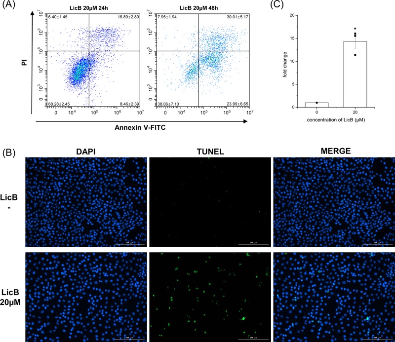
LicB causes apoptosis of HaCaT cells. (A) Dot plot of Annexin V-FITC/PI double staining over time in HaCaT cells treated with 20 μM LicB. (B) TUNEL observed with Cytation5 in HaCaT cells. (C) Proportion of TUNEL-positive cells in the LicB-treated group compared with the control group. The rate of apoptosis and necrosis obtained from each experiment (n = 4) and the proportion of TUNEL-positive cells (n = 3) are presented as the mean ± standard error. Statistical significance was evaluated by t-test. *p < 0.05.
The activity of various apoptosis-related factors was detected to investigate the apoptosis mechanism caused by LicB. The active forms of PARP and caspase-3, which are commonly activated in the apoptosis pathway, were detected (Fig. 4A) and they significantly increased in the 20 μM treatment group (Fig. 4B). The ratio of Bax/Bcl-2, a key marker of intrinsic apoptosis, and cleaved caspase-8, a marker of extrinsic apoptosis, increased concentration-dependently. These results indicated that LicB activated both the intrinsic and extrinsic pathways of apoptosis (Fig. 4C and D).
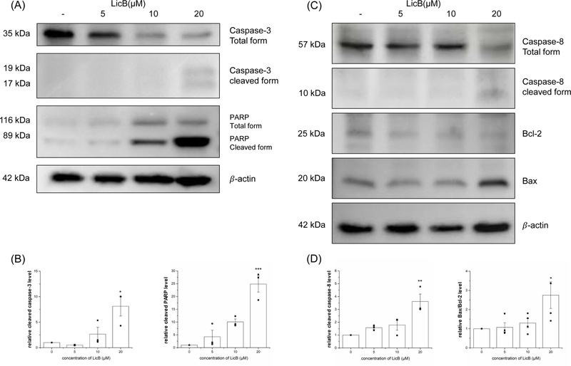
Enhanced apoptosis factors related to intrinsic and extrinsic pathways by LicB. The HaCaT cells were stimulated by various concentrations of LicB (0, 5, 10, and 20 μM) for 24 h. (A) Caspase-3 and PARP band. (B) Quantification of cleaved caspase-3 and cleaved PARP band and comparison of their expression with the controls. (C) Caspase-8, Bcl-2, and Bax band. (D) Quantification of cleaved caspase-8 and Bax/Bcl-2 band and comparison of their expression with the controls. The graph shows the values obtained from three independent experiments (n = 3), and statistical significance was evaluated via one-way ANOVA. *p < 0.05, **p < 0.01, ***p < 0.001.
Under inflammatory conditions induced by TNF-α and IFN-γ, low concentrations of LicB modestly suppressed the expression of chemokines associated with immune cell recruitment, including Th2 cells, eosinophils, and neutrophils,17,18 while the expression of major pro-inflammatory cytokines remained largely unaffected. Although inflammatory signaling pathways such as NF-κB and JAK-STAT have been implicated in the anti-inflammatory effects of chalcone derivatives,3,4,19,20 these mechanisms were not directly examined in the present study as the anti-inflammatory effects of LicB were considered mild. In addition, the anti-inflammatory effects were evaluated at the mRNA level in the present study; further validation at the protein level would strengthen these findings. Conversely, exposure to relatively higher concentrations of LicB resulted in a significant reduction in cell viability and a marked increase in apoptotic cell death. However, the same concentration of LicB did not significantly affect cell viability in hepatocyte and hepatoma cell lines, AML12 and Huh7, suggesting that keratinocytes may be more susceptible to LicB. Furthermore, LicB has been reported to exert protective effects against tissue injury, oxidative stress, and various inflammatory models within a similar concentration range (10–20 μM).14,15,21 These data indicate that LicB may induce cytotoxicity through apoptosis at relatively low concentrations in skin cells. Given the essential role of keratinocytes in maintaining epidermal integrity and barrier function, such cytotoxic effects may have detrimental consequences for skin homeostasis. While licorice toxicity has mainly been discussed in the context of oral exposure, our results highlight a previously underexplored potential for skin-related toxicity of individual licorice-derived components. Although evaluated in isolation in this study, these findings underscore the importance of assessing dose-dependent safety and cytotoxic effects of natural product–derived compounds. In this regard, as this study utilized a single immortalized keratinocyte cell line (HaCaT), further studies using primary human keratinocytes or complementary in vivo models would help validate the relevance of these findings.
In conclusion, our findings provide a new perspective on the safety of natural product components by suggesting that LicB, a licorice-derived compound often associated with beneficial biological activities, may induce cytotoxicity at relatively low concentrations in keratinocytes. Notably, concentrations considered pharmacologically relevant were associated with apoptosis in keratinocytes, indicating a previously underappreciated safety concern in skin cells. These results highlight the importance of cell-type-specific toxicity evaluation of natural product-derived compounds, particularly in the context of skin exposure.
Acknowledgments
This work was supported by the National Research Foundation of Korea (RS-2023-00251463). This research was supported by Global-Learning & Academic research institution for Master’s·PhD students, and Postdocs (LAMP) Program of the National Research Foundation of Korea (NRF) grant funded by the Ministry of Education (No. RS-2024-00442775).
Conflict of Interest
The authors declare that they have no conflicts of interest.
References
-
Shaikh, S.; Lee, E. J.; Ahmad, K.; Choi, I. Front. Mol. Biosci. 2024, 11, 1440132.
[https://doi.org/10.3389/fmolb.2024.1440132]

-
Husain, I.; Bala, K.; Khan, I. A.; Khan, S. I. Food Front. 2021, 2, 449–485.
[https://doi.org/10.1002/fft2.110]
-
Furusawa, J.-I.; Funakoshi-Tago, M.; Mashino, T.; Tago, K.; Inoue, H.; Sonoda, Y.; Kasahara, T. Int. Immunopharmacol. 2009, 9, 499–507.
[https://doi.org/10.1016/j.intimp.2009.01.031]
-
Du, Z.; Liao, X.; Kang, Q. Mol. Cell. Toxicol. 2025, 21, 197–203.
[https://doi.org/10.1007/s13273-024-00437-7]
-
Huang, Z.; Jin, G. Biol. Pharm. Bull. 2022, 45, 730–737.
[https://doi.org/10.1248/bpb.b21-00991]
-
Oh, H.; Yoon, G.; Shin, J.-C.; Park, S.-M.; Cho, S.-S.; Cho, J. H.; Lee, M.-H.; Liu, K.; Cho, Y. S.; Chae, J.-I.; Shim, J.-H. Int. J. Oncol. 2016, 48, 1749–1757.
[https://doi.org/10.3892/ijo.2016.3365]
-
Omar, H. R.; Komarova, I.; El-Ghonemi, M.; Fathy, A.; Rashed, R.; Abdelmalak, H. D.; Yerramadha, M. R.; Ali, Y.; Helal, E.; Camporesi, E. M. 2012, 3, 125–138.
[https://doi.org/10.1177/2042018812454322]
-
Calò, L. A.; Zaghetto, F.; Pagnin, E.; Davis, P. A.; Mozzi, P. D.; Sartorato, P.; Martire, G.; Fiore, C.; Armanini, D. J. Clin. Endocrinol. Metab. 2004, 89, 1973–1976.
[https://doi.org/10.1210/jc.2003-031545]
-
Stewart, P. M.; Wallace, A. M.; Valentino, R.; Burt, D.; Shackleton, C. H.; Edwards, C. R. Lancet 1987, 2, 821–824.
[https://doi.org/10.1016/S0140-6736(87)91014-2]
-
Tsai, J. P.; Hsiao, P. C.; Yang, S. F.; Hsieh, S. C.; Bau, D. T.; Ling, C. L.; Pai, C. L.; Hsieh, Y. H. PLoS One 2014, 9, e86537.
[https://doi.org/10.1371/journal.pone.0086537]
-
Yang, G.; Lee, H. E.; Yeon, S. H.; Kang, H. C.; Cho, Y.-Y.; Lee, H. S.; Zouboulis, C. C.; Han, S.-H.; Lee, J.-H.; Lee, J. Y. Phytother. Res. 2018, 32, 2551–2559.
[https://doi.org/10.1002/ptr.6195]
-
D'Arcy, M. S. Cell Biol. Int. 2019, 43, 582–592.
[https://doi.org/10.1002/cbin.11137]
-
Anderton, H.; Alqudah, S. Biochem. J. 2022, 479, 1621–1651.
[https://doi.org/10.1042/BCJ20210606]
-
Li, Q.; Feng, H.; Wang, H.; Wang, Y.; Mou, W.; Xu, G.; Zhang, P.; Li, R.; Shi, W.; Wang, Z.; Fang, Z.; Ren, L.; Wang, Y.; Lin, L.; Hou, X.; Dai, W.; Li, Z.; Wei, Z.; Liu, T.; Wang, J.; Guo, Y.; Li, P.; Zhao, X.; Zhan, X.; Xiao, X.; Bai, Z. EMBO Rep. 2022, 23, e53499.
[https://doi.org/10.15252/embr.202153499]
-
Wu, Y.; Feng, K.; Chen, Y.; Zhang, H.; Zhang, M.; Han, B.; Chen, X.; Yang, L.; Wang, X.; Li, W.; Tang, J. J. Ethnopharmacol. 2024, 335, 118600.
[https://doi.org/10.1016/j.jep.2024.118600]
- Gao, X.-P.; Qian, D.-W.; Xie, Z.; Hui, H. Iran. J. Basic Med. Sci. 2017, 20, 131–137.
-
Jahnz-Rozyk, K.; Targowski, T.; Paluchowska, E.; Owczarek, W.; Kucharczyk, A. Allergy 2005, 60, 685–688.
[https://doi.org/10.1111/j.1398-9995.2005.00774.x]
-
Kim, E. J.; Park, H.; Kim, E.-Y.; Kim, D. K.; Jung, H.-S.; Sohn, Y. Life Sci. 2024, 344, 122560.
[https://doi.org/10.1016/j.lfs.2024.122560]
-
Song, M.; Yoon, G.; Choi, J.-S.; Kim, E.; Liu, X.; Oh, H.-N.; Chae, J.-I.; Lee, M.-H.; Shim, J.-H. Phytother. Res. 2020, 34, 2032–2043.
[https://doi.org/10.1002/ptr.6661]
-
Yadav, V. R.; Prasad, S.; Sung, B.; Aggarwal, B. B. Int. Immunopharmacol. 2011, 11, 295–309.
[https://doi.org/10.1016/j.intimp.2010.12.006]
-
Kim, A.; Kim, Y. R.; Jayasinghe, J. N. C.; Park, M.; Cha, S.; Park, S.-M.; Kim, N. S. Free Radic. Biol. Med. 2025, 241, 204–219.
[https://doi.org/10.1016/j.freeradbiomed.2025.09.022]