
Chemical Components and Biological Effects of Ethanol Extract from Curcuma vinhlinhensis
Abstract
Curcuma vinhlinhensis is a recent novel species in Vietnam. In this study, the chemical composition of ethanol extract of leaf and rhizome from C. vinhlinhensis was first analyzed by gas chromatography/mass spectrometry and then 13 and 22 chemical compounds were found to be present in the two extracts, respectively. The ethanol leaf extract contained 36.57% of (E)-labda-8(17),12-diene-15,16-dial, 13.17% of octacosane, 8.63% of n-hexadecanoic acid, 7.16% of neophytadiene, 6.55% hexacosane, and other compounds in small amount (< 5%). Meanwhile, the ethanol rhizome extract contained predominantly (E)-labda-8(17),12-diene-15,16-dial (64.82%), copaene (5.33%), and other compounds (< 5%). Both extracts from leaf and rhizome displayed antibacterial activity against tested bacterial pathogens including Bacillus cereus, Staphylococcus aureus, Staphylococcus saprophyticus, Escherichia coli, Enterobacter hormaechei, Klebsiella pneumoniae, Salmonella typhimurium, and Shigella flexneri. The antioxidant property displayed by radical scavenging activity on DPPH and ABTS has also been reported. The IC50 value of DPPH radical scavenging activity was 729.88 ± 46.54 μg/mL and 1666.01 ± 134.16 μg/mL for rhizome extract and leaf extract, respectively. Additionally, IC50 value of ABTS radical scavenging activity was 257.04 ± 13.89 μg/mL for rhizome extract and 424.61 ± 24.721 μg/mL for leaf extract. These results have shown that the ethanol extract of C. vinhlinhensis could potentially be used for pharmaceutical applications and product development.
Keywords:
Antibacterial activity, Antioxidant activity, Chemical composition, Curcuma vinhlinhensis, Ethanol extractIntroduction
Curcuma belongs to the Zingiberaceae family, which is known as a group of popular herbs in dietary gastronomy and traditional medicine in Asian countries such as China, Korea, Japan, Thailand, In Vietnam, 29 species of Curcuma have been recorded up to date.1 Studies on the biological activity of various Curcuma plants have been conducted and published. Curcuma comosa Roxb. ethanol extract showed the protective activity against cisplatin-induced nephrotoxicity in mice. Cisplatin is a widely used chemotherapeutic agent for the treatment of various malignant tumors.2 Curcuma phaeocaulis Valeton ethanolic extract displayed anti-tumor potential for breast cancer cells.3 Ethanolic extract of C. mangga and its chloroform and hexane fractions showed analgesic and anti-inflammatory activities,4 and similar results were also observed for ethanolic extract of C. zedoaria.5 Antibacterial and antifungal activities were found in various extracts of C. zedoaria and C. malabarica tubers.6 Ethanolic extract of Curcuma longa rhizome was shown to possess antioxidant and antidiabetic capability.7 Essential oils, ethanolic extract from C. longa exhibited larvicidal and biting deterrent activity against yellow fever, dengue, malaria vectors such as Aedes aegypti and Anopheles quadrimaculatus.8 In addition, the amoebicidal activity of C. longa ethanol extract was also observed when the extract showed significant inhibition on Acanthamoeba cyst multiplications.9 Moreover, Curcumin, a common phenolic compound in Curcuma, is known for many outstanding biological effects such as antioxidant activities, cancer prevention, brain aging and neurodegenerative protection,10–13 acting against retardation of wound healing by aspirin.14 The compound xanthorrhizol, isolated from C. xanthorrhiza Roxb., has been confirmed for antimicrobial, antioxidant, anti-inflammatory, anticancer, antihyperglycemic, and antihypertensive, antiplatelet, nephroprotective, and hepatoprotective properties.15,16 With valuable biological properties, xanthorrizol from C. xanthorrhiza Roxb. interested and applied research in the pharmaceutical industry.
Curcuma vinhlinhensis is one of two new species belonging to the Curcuma subgen. Ecomata Zingiberaceae: Zingibereae was recently discovered in Vietnam, Quang Tri Province, Vinh Linh Commune.17 Chemical composition and biological activities of this new species, therefore, have not been investigated to date. In this study, ethanol extract of C. vinhlinhensis leaves and rhizomes, for the first time, were examined for chemical composition, antibacterial, and antioxidant activity, providing novel insights into the Curcuma genus and the potential practical applications of this herb.
Materials and Methods
Plant materials – Curcuma vinhlinhensis plants were collected from Vietnam, Da Nang City, Duy Xuyen Commune on 10 December 2022, at coordinates 15o 49ʹ 39.62ʺ N; 108o 11ʹ 30.75ʺ E (Fig. 1). The voucher specimens, NPN_QN_35 and NPN_QN_36, were deposited at the Herbarium of Faculty of Biology and Biotechnology, University of Science, Vietnam National University Ho Chi Minh City. The scientific name of the studied species was determined using a comparative morphological method. The reproductive and vegetative organs were thoroughly examined and compared with those described in previously published studies on the genus Curcuma.1,17 The rhizome and leaves samples were grounded into powder and subsequently used for ethanol extraction.
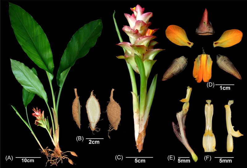
Curcuma vinhlinhensis D. D. Nguyen & T. A. Le. Habit (A), Root tubers (B), Inflorescences (C), Flower dissection with labellum, two lateral staminodes, dorsal corolla lobe and two lateral corolla lobes (D), Floral tube with anther (E) and Anther in front and side view (F). Photos: Nga Nguyen-Phi
Bacterial strains and culture condition – Nine strains from American Type Culture Collection (ATCC) were used to determine the antibacterial activity of ethanolic extracts. These strains include four Gram-positive bacteria such as Bacillus cereus (ATCC 11774), Staphylococcus aureus (ATCC 29213), Staphylococcus aureus (ATCC 25923), Staphylococcus saprophyticus (ATCC BAA-750), and five Gram-negative bacteria, such as Escherichia coli (ATCC 25922), Enterobacter hormaechei (ATCC 700323), Klebsiella pneumoniae (ATCC 13883), Salmonella typhimurium (ATCC 13311), and Shigella flexneri (ATCC 12022). All strains were stored at −80℃ and pre-cultured in Luria-Bertani broth at 37℃ for 16 h before further use.
Preparation of ethanolic extracts – One hundred grams of rhizome and leaf dry powder were soaked in 500 mL of ethanol 99% (Thermo Fisher Scientific, USA) for 72 hours at room temperature. The extracts were filtered 3 times through Whatman papers and concentrated using a rotary evaporator under reduced pressure at 50℃ to obtain ethanol-free concentrated brown extracts.18 These extracts were used for analysis of chemical composition and antibacterial activity.
Analysis of chemical profile – Chemical components of ethanolic extracts were analyzed using TRACE 1310 Gas Chromatograph (Thermo Fisher Scientific, Waltham, MA, USA) coupled to a ISQ 7000 single quadrupole mass spectrometer. DB-5MS column (30 m × 0.25 mm × 0.25 μm) was used as the stationary phase and Helium was used as the carrier gas with a flow rate 1.2 mL/min. Samples were injected with split ratio of 30:1, 1 min splitless time, flow rate 36 mL/min, at 250℃. The electron impact ionization was set as 70 eV and the filament source temperature was set at 250℃. The oven temperature was set at 80℃ for 5 min then increased 20℃/min until 280℃ was reached, and finally held at 280℃ for 10 min. The acquisitions scan mass range of MS was 29–650 m/z with scanning frequency of 2 scans/sec. Chemical composition of the samples was determined by comparing the mass spectra of the samples to those from the NIST 2017 library.
Antibacterial activity assay – Antibacterial activity of ethanolic extracts was determined using disc diffusion method. Bacterial strains were grown in Luria-Bertani broth at 37℃ until culture turbidity reached 0.5 McFarland equivalent and were subsequently spread on Mueller Hinton agar plates with depth of ~ 4 mm. Ten microliter (10 μL) of ethanolic extracts (100 mg/mL) were load into the discs (diameter of 6 mm) and the plates were incubated at 37℃ for 16–18 hours. Antimicrobial activity against nine bacterial strains was determined based on growth inhibition zones. Gentamicin (10 μg, Nam Khoa BioTek, Vietnam) was used as positive control.
DPPH radical scavenging assay – Free radical scavenging ability of the extracts was evaluated using DPPH (2,2-diphenyl-1-picrylhydrazyl) radical scavenging assay.8 Equal amount (100 μL) of sample and DPPH solution (300 μM) were mixed, incubated in the dark for 30 min at room temperature, and then OD517 was measured by UV/vis spectrophotometer (UVS 2800, Labome, USA). DPPH radical scavenging activity (DPPHRSA) of the extracts were calculated using the following formula:
DPPHRSA (%) = (Abscontrol − Abssample)/Abscontrol × 100%
where Abscontrol is the absorbance of the DPPH solution in ethanol while Abssample is the absorbance of the DPPH solution-ethanol extract mixture. The IC50 value was estimated from the concentration-response curve of antioxidant activity obtained from the extracts. The results were compared with ascorbic acid as the reference standard.
ABTS radical scavenging assay – The ABTS (2,2'-azino-bis (3-ethylbenzothiazoline-6-sulfonic acid)) radical scavenging ability of the extracts were evaluated using Maeng’s protocol.19 Firstly, solution A was prepared by gently mixing equal amount of 7.0 mM ABTS and 2.45 mM K2S2O8 of the sample and incubated in the dark 18 hours at room temperature. Secondly, 0.1 mL of each extract was gently mixed with 3 mL solution A, brought up to 5 mL by ethanol. The mixture was incubated in the dark for 15 minutes at room temperature and subsequently measured for OD734. Ascorbic acid was used as reference standard and the standard curve (0–15 ppm) was constructed with the equation y = −0.0278x + 0.421, R2 = 0.9990, where y is the absorbance at 734 nm, and x is the sample concentration (μg/mL). The sample concentration was calculated from the standard curve equation and expressed as μg/mL ascorbic acid.
Data analysis – Antimicrobial assay was performed in triplicate, while antioxidant activity tests were conducted six times for experiments involving plant extracts and at least three times for the standard control using ascorbic acid. Data were presented as mean ± standard deviation (SD). Statistical comparisons were conducted with Statgraphics Centurion 15 using one-way ANOVA. Mean differences between extracts (leaf vs. rhizome) and positive controls for each microbial strain were identified using Fisher’s LSD test (p < 0.05).
Results and Discussion
The results of GC-MS analysis showed that a total of 13 and 22 compounds were detected in the profile of ethanol leaf extracts and rhizome ethanol extract, respectively (Table 1, Fig. 2). The presence of (E)-Labda-8(17),12-diene-15,16-dial was predominant in both of the extracts from leaf (36.57%) and rhizome (64.82%). Previous studies have reported the presence of this compound in rhizome samples of some Curcuma species and other Zingiberaceae plants such as C. comosus var. bakeri, C. rubescens, C. aeruginosa, Haemodorum brevicaule, Alpinia oxyphylla, and A. pumila, while there is limited data on the presence of this compound in leaf samples.20–24 The content of (E)-Labda-8(17),12-diene-15,16-dial depends on Curcuma species, from 11.45% in C. rubescens and 14.58% in C. cotuana to 33.37% in C. thorelii and up to 94.17% in C. aeruginosa.23,24 This compound was also found in as major constituent in C. amada while it appears as a minor component in C. mangga.25,26

Chemical profile of C. vinhlinhensis ethanol extracts
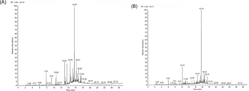
The GC chromatogram of the ethanol extracts from C. vinhlinhensis. (A) Leaf and (B) Rhizome.
Furthermore, the biological activities of (E)-labda-8(17),12-diene-15,16-dial and its derivatives have been documented. Previous studies have demonstrated their potent antibacterial and antioxidant effects, alongside the suppression of nitric oxide (NO) production and the down-regulation of inducible nitric oxide synthase (iNOS) mRNA expression levels.26–28
However, antioxidant and antibacterial activities of (E)-labda-8(17),12-diene-15,16-dial and its derivatives in C. vinhlinhensis remain to be fully elucidated. Consequently, further investigation is required to isolate this compound from C. vinhlinhensis and focus on correlating chemical compound with specific bioactivities to confirm its therapeutic efficacy in this species.
Linolenic acid, neophytadiene, n-hexadecanoic acid, octacosane were also found to be present in high amount in ethanol leaf extract (7.09%–13.17%) while only n-hexadecanoic acid, with very low amount (1.94%), was found in ethanol rhizome extract. Beside (E)-Labda-8(17),12-diene-15,16-dial, the remaining 21 compounds accounted for only 0.18%–5.33% in the ethanol rhizome extract. The diversity of the composition as well as the accumulation of chemical compounds in different ethanolic extracts from C. vinhlinhensis, therefore, could provide the basis for further exploration of this new species’ potential bioactivities.
The results from disc diffusion assay showed that both ethanol extracts from leaf and rhizome had inhibitory effect against 9 tested bacterial strains with large inhibition zone from 15.33 ± 2.08 mm to 29.17 ± 2.36 mm (Table 2). Except for E. hormachei ATCC700323 and S. aureus ATCC 25923, no difference in antibacterial activity on the other strains between leaf and root samples has been observed. The positive effect of both extracts was most pronounced on K. pneumoniae ATCC 13883 and the two most common reference S. aureus strains used for antibacterial tests. The leaf and rhizome extracts produced inhibition zones against S. aureus ATCC 29213, a weak β-lactamase producer, measuring 29.17 ± 2.36 mm and 26.33 ± 2.08 mm, respectively. Furthermore, the rhizome extract demonstrated a stronger inhibitory effect against the β-lactamase-non-producing S. aureus ATCC 25923, with an inhibition zone of 26.33 ± 1.53 mm, compared to the leaf extract (21.83 ± 2.02 mm).
Antibacterial activity of C. vinhlinhensis ethanol extracts
The antibacterial activity of ethanol extracts from other Curcuma species has also been reported. The ethanol extract from C. malabarica showed inhibitory effect on S. aureus, B. subtilis, Micrococcus luteus, Proteus mirabilis, K. pneumoniae, while the ethanol extract from C. zedoaria species had no effect on S. aureus. The ethanol extracts of these two species also did not inhibit the growth of E. coli.6 Additionally, the antibacterial activity of C. longa ethanol extract was displayed in E. coli, S. aureus, Vibrio cholerae, Streptococcus pyogenes, S. agalactiae, but not in S. typhimurium, S. enteritidis, and L. monocytogenes.29,30 Similarly, in this study, the antibacterial activity of both ethanol extracts from C. vinhlinhensis showed a broad antibacterial spectrum and the inhibition of E. coli could be explained by the presence of the main compound (E)-Labda-8(17),12-diene-15,16-dial.31
Ethanol extracts from both C. vinhlinhensis rhizome and leaf exhibited DPPH radical scavenging activity, of which the extract concentration was proportional to radical scavenging activity (Fig. 3A). Free radical scavenging ability of the rhizome extract was shown to be more than 2-folds higher than that of the leaf extract with IC50 of 729.88 ± 46.54 μg/mL compared with 1666.01 ± 134.16 μg/mL in case of the leaf extract (Fig. 3B). The DPPH radical scavenging ability of ethanol extracts from other Curcuma species have also been reported in previous studies with IC50 value of C. longa from 27.2 μg/mL to 34.86 μg/mL.7,32 or from 26.8 ± 1.4 μg/mL to 765.0 ± 39.3 μg/mL in the case of C. zedoaria, C. xanthorrhiza, C. aeruginosa, and C. mangga.33
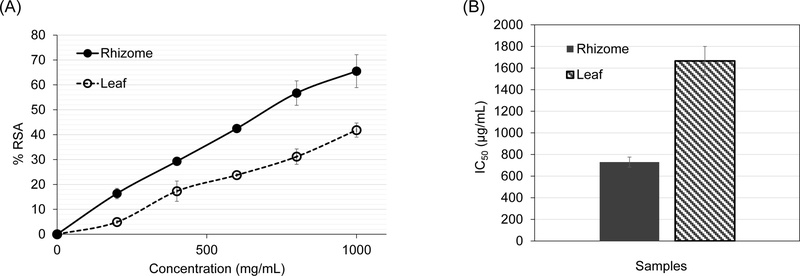
Determination of a DPPH radical scavenging activity (%RSA) in various concentrations of ethanol extracts (A) and IC50 of radical scavenging activity (B). Results expressed as mean ± SD of six replicates.
Fig. 4A further confirmed the antioxidant activity of the two ethanol extracts from C. vinhlinhensis via ABTS radical scavenging activity which was found to be proportional to the extract concentration. Additionally, IC50 value was also calculated with 257.04 ± 13.89 μg/mL of the rhizome extract and 424.61 ± 24.721 μg/mL of the leaf extract (Fig. 4B), which means that antioxidant activity of the rhizome extract was about 1.6 folds higher. Despite of that, this activity of C. vinhlinhensis extracts was relatively 1.9–3.2 folds higher than that of the C. longa 70% ethanol extract (840 ± 30 μg/mL) in the study of Kim et al..34
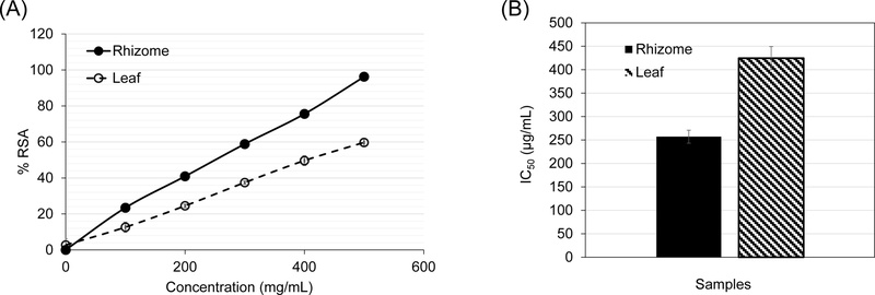
Determination of an ABTS radical scavenging property (%RSA) in various concentrations of ethanol extracts (A) and IC50 of radical scavenging activity (B). Results expressed as mean SD of six replicates.
In conclusion, the Curcuma genus has long been used as health food products and pharmaceutical products. As a result, chemical composition and biological activities of Curcuma species have been increasingly studied. This study, for the first time provides a detailed chemical profile as well as biological activities of the absolute ethanolic extracts of the rhizome and leaf of C. vinhlinhensis, a newly found Curcuma species in Central Vietnam. The data showed besides of the major compound (E)-Labda-8(17),12-diene-15,16-dial in both extracts, chemical composition and concentration were very different between the two. However, the two ethanolic extracts had, even though different, broad-spectrum antibacterial activity against both Gram-positive and Gram-negative bacteria. The two extracts exhibited antioxidant activity, as demonstrated by their ability to scavenge DPPH and ABTS radicals; however, their antioxidant potency remains modest compared with ascorbic acid, with the rhizome ethanolic extract showing comparatively stronger activity. Therefore, further investigations are required to more accurately evaluate their potential for pharmaceutical and related applications. Specifically, future studies should address cytotoxicity against cancer cell lines, determine minimum inhibitory and bactericidal concentrations (MIC/MBC), and conduct comprehensive safety assessments.
Acknowledgments
This work was supported by Industrial University of Ho Chi Minh City.
Conflicts of Interest
The authors declare that they have no conflicts of interest.
References
- Nguyen, Q. B.; Nguyen, P. H.; Nghiem, D. T.; Do, H. C.; Nguyen, T. T. VNU J. Sci. 2019, 35, 102–107.
-
Jariyawat, S.; Kigpituck, P.; Suksen, K.; Chuncharunee, A.; Chaovanalikit, A.; Piyachaturawat, P. J. Nat. Med. 2009, 63, 430–436.
[https://doi.org/10.1007/s11418-009-0345-5]

-
Chen, X.; Pei, L.; Zhong, Z.; Guo, J.; Zhang, Q.; Wang, Y. Phytomedicine 2011, 18, 1238–1243.
[https://doi.org/10.1016/j.phymed.2011.06.017]
-
Ruangsang, P.; Tewtrakul, S.; Reanmongkol, W. J. Nat. Med. 2010, 64, 36–41.
[https://doi.org/10.1007/s11418-009-0365-1]
-
Ullah, H.; Zaman, S.; Juhara, F.; Akter, L.; Tareq, S. M.; Masum, E. H.; Bhattacharjee, R. BMC Complement. Altern. Med. 2014, 14, 1–12.
[https://doi.org/10.1186/1472-6882-14-346]
-
Wilson, B.; Abraham, G.; Manju, V. S.; Mathew, M.; Vimala, B.; Sundaresan, S.; Nambisan, B. J. Ethnopharmacol. 2005, 99, 147–151.
[https://doi.org/10.1016/j.jep.2005.02.004]
-
Sabir, S. M.; Zeb, A.; Mahmood, M.; Abbas, S.; Ahmad, Z.; Iqbal, N. Braz. J. Biol. 2020, 81, 737–740.
[https://doi.org/10.1590/1519-6984.230628]
-
Ali, A.; Wang, Y.‐H.; Khan, I. A. J. Med. Entomol. 2015, 52, 979–986.
[https://doi.org/10.1093/jme/tjv072]
-
El‐Sayed, N. M.; Ismail, K. A.; Ahmed, S. A.‐E.‐G.; Hetta, M. H. Parasitol. Res. 2012, 110, 1985–1992.
[https://doi.org/10.1007/s00436-011-2727-3]
-
Forouzanfar, F.; Read, M. I.; Barreto, G. E.; Sahebkar, A. IUBMB Life 2020, 72, 652–664.
[https://doi.org/10.1002/iub.2209]
-
Scapagnini, G.; Caruso, C.; Calabrese, V. Adv. Exp. Med. Biol. 2010, 698, 27–35.
[https://doi.org/10.1007/978-1-4419-7347-4_3]
-
Jovanovic, S. V.; Boone, C. W.; Steenken, S.; Trinoga, M.; Kaskey, R. B. J. Am. Chem. Soc. 2001, 123, 3064–3068.
[https://doi.org/10.1021/ja003823x]
-
Rajakrishnan, V.; Viswanathan, P.; Rajasekharan, K. N.; Menon, V. P. Phytother. Res. 1999, 13, 571–574.
[https://doi.org/10.1002/(SICI)1099-1573(199911)13:7<571::AID-PTR494>3.3.CO;2-Z]
-
Pawar, R. S.; Toppo, F. A.; Mandloi, A. S.; Shaikh, S. Indian J. Pharmacol. 2015, 47, 160–166.
[https://doi.org/10.4103/0253-7613.153422]
-
Oon, S. F.; Nallappan, M.; Tee, T. T.; Shohaimi, S.; Kassim, N. K.; Sa’ariwijaya, M. S. F.; Cheah, Y. H. Cancer Cell Int. 2015, 15, 1–15.
[https://doi.org/10.1186/s12935-015-0255-4]
-
Simamora, A.; Timotius, K. H.; Yerer, M. B.; Setiawan, H.; Mun'im, A. Phytomedicine 2022, 105, 154359.
[https://doi.org/10.1016/j.phymed.2022.154359]
-
Nguyen, D. D.; Le, T. A.; Hoang, Q. H.; Le, Q. T.; Nguyen, E. Biodiversitas 2022, 23, 2512–2519.
[https://doi.org/10.13057/biodiv/d230531]
-
Le, H. T.; Luu, T. N.; Nguyen, H. M. T.; Nguyen, D. H.; Le, P. T. Q.; Trinh, N. N.; Nguyen, H. D.; Van, H. T. Plant Sci. Today 2021, 8, 273–277.
[https://doi.org/10.14719/pst.2021.8.2.1086]
-
Maeng, J.‐H.; Muhammad Shahbaz, H.; Ameer, K.; Jo, Y.; Kwon, J.‐H. J. Food Process Eng. 2017, 40, e12421.
[https://doi.org/10.1111/jfpe.12421]
-
Awin, T.; Mediani, A.; Maulidiani; Leong, S. W.; Faudzi, S. M. M.; Shaari, K.; Abas, F. J. Food Compos. Anal. 2019, 77, 66–76.
[https://doi.org/10.1016/j.jfca.2019.01.004]
-
Otsuka, S.; Kawamura, M.; Fujino, S.; Nakamura, F.; Arai, D.; Fusetani, N.; Nakao, Y. J. Agric. Food Chem. 2022, 70, 3300–3309.
[https://doi.org/10.1021/acs.jafc.2c00020]
-
Jalaja, R.; Leela, S. G.; Mohan, S.; Nair, M. S.; Gopalan, R. K.; Somappa, S. B. Bioorg. Chem. 2021, 108, 104664.
[https://doi.org/10.1016/j.bioorg.2021.104664]
-
Peng, W.; Li, P.; Ling, R.; Wang, Z.; Feng, X.; Liu, J.; Yang, Q.; Yan, J. Molecules 2022, 27, 565.
[https://doi.org/10.3390/molecules27020565]
- Van, H. T.; Tran, M. D.; Tran, T. K. N.; Nguyen, H. T. D.; Nguyen, N. A.; Huynh, N. T. A.; Nguyen, Q. H.; Le, T. T.; Nguyen, P. N.; Pham, T. V. Plant Sci. Today 2023, 10, 83–89.
-
Alan Sheeja, D. B. A.; Nair, M. S. Biochem. Syst. Ecol. 2012, 44, 264–266.
[https://doi.org/10.1016/j.bse.2012.06.008]
-
Liu, Y.; Nair, M. G. Food Chem. 2011, 124, 527–532.
[https://doi.org/10.1016/j.foodchem.2010.06.064]
-
Ghosh, S.; Indukuri, K.; Bondalapati, S.; Saikia, A. K.; Rangan, L. Eur. J. Med. Chem. 2013, 66, 101–105.
[https://doi.org/10.1016/j.ejmech.2013.05.034]
-
Trang, N. M.; Vinh, L. B.; Phong, N. V.; Yang, S. Y. Planta Med. 2025, 91, 29–39.
[https://doi.org/10.1055/a-2440-5166]
-
Marasini, B. P.; Baral, P.; Aryal, P.; Ghimire, K. R.; Neupane, S.; Dahal, N.; Singh, A.; Ghimire, L.; Shrestha, K. BioMed Res. Int. 2015, 2015, 265425.
[https://doi.org/10.1155/2015/265425]
-
Thongson, C.; Davidson, P. M.; Mahakarnchanakul, W.; Vibulsresth, P. J. Food Prot. 2005, 68, 2054–2058.
[https://doi.org/10.4315/0362-028X-68.10.2054]
- Sheeja, A. D. B.; Nair, M. S. Indian J. Chem. Sect. B 2014, 53, 319–324.
-
Ak, T.; Gülçin, İ. Chem. Biol. Interact. 2008, 174, 27–37.
[https://doi.org/10.1016/j.cbi.2008.05.003]
-
Awin, T.; Mediani, A.; Maulidiani; Shaari, K.; Faudzi, S. M. M.; Sukari, M. A. H.; Lajis, N.; Abas, F. Ind. Crop. Prod. 2016, 94, 342–352.
[https://doi.org/10.1016/j.indcrop.2016.08.020]
-
Kim, G.‐H.; Duan, Y.; Kim, H.‐S. J. Korean Oil Chemists. Soc. 2016, 33, 521–528.
[https://doi.org/10.12925/jkocs.2016.33.3.521]