
Ameliorative Effects of Lonicera japonica Extract on Scopolamine-Induced Memory Deficits via Cholinergic Modulation
Abstract
Alzheimer’s disease (AD) is a progressive neurodegenerative disorder characterized by memory and learning decline leading to dementia. Lonicera japonica (LJ), traditionally used in East Asian medicine, has shown various pharmacological effects including anti-inflammatory, antioxidant, and antimicrobial activities. Previous studies demonstrated that LJ extract protects HT22 neuronal cells against glutamate-induced neurotoxicity. In this study, we examined the cognitive-enhancing effects of LJ using a scopolamine-induced memory impairment model in mice. Spatial and associative learning were assessed using the Morris water maze and passive avoidance tests, respectively. LJ extract was prepared via ultrasonic methanolic extraction and administered orally at 100, 200, and 300 mg/kg body weight. Results showed that LJ significantly improved scopolamine-induced memory deficits and inhibited acetylcholinesterase activity. These findings suggest that LJ may enhance cognitive function through modulation of the cholinergic system and support its potential as a candidate for nutraceutical development targeting memory impairment.
Keywords:
Lonicera japonica, Alzheimer’s disease, Cognitive enhancing activity, Acetylcholine esteraseIntroduction
Dementia is a complex neurological disorder characterized by progressive impairments in multiple cognitive domains, including memory, language, and executive functions, resulting from widespread brain dysfunction.1 Alzheimer’s disease (AD) represents the most common form of dementia, accounting for approximately 50–60% of cases.2 Its key pathological features include amyloid-β plaque accumulation, tau hyperphosphorylation with neurofibrillary tangle formation, and gradual neuronal loss associated with cellular stress responses.3,4
Although considerable research has been conducted, the exact cause of AD has not been fully elucidated, which limits the development of disease-modifying therapies.5 In the early stage, symptoms are often mild and difficult to detect, but disease progression leads to extensive neuronal degeneration across multiple brain regions, resulting in severe cognitive and functional decline.6 Currently approved drugs, such as Donepezil, Rivastigmine, and Galantamine, mainly offer symptomatic relief, highlighting the need for safer and more effective treatment strategies.7,8
For the assessment of cognitive function in experimental studies, behavioral tests such as the Morris water maze (MWM) and the passive avoidance test are commonly used. The MWM is designed to evaluate spatial learning and memory,9 whereas the passive avoidance test is used to assess both short-term and long-term memory retention.10
Lonicera japonica (Caprifoliaceae) is a perennial flowering plant that is widely distributed throughout East Asia, especially in the temperate climates of China, Korea, and Japan, where it grows naturally in fields, forest edges, and mountainous regions.11,12 For centuries, it has been an important component of traditional East Asian medicine and is commonly described as having “heat-clearing” and “detoxifying” properties, being frequently prescribed for the management of febrile illnesses and various infectious diseases.13
In recent decades, scientific interest in L. japonica has increased significantly, and numerous studies have reported a broad range of pharmacological activities. These include antibacterial and antiviral effects, along with protective actions on the liver and regulatory effects on lipid metabolism, such as antihyperlipidemic activity.14–16 In addition, it has been consistently shown to exert strong antioxidant capacity and to suppress inflammatory responses, suggesting a role in mitigating oxidative stress- and inflammation-related disorders.17–19
Phytochemical analyses have further identified that L. japonica is rich in diverse bioactive compounds. Major constituents include flavonoids, iridoid glycosides, and phenolic acids such as chlorogenic acid, all of which are believed to contribute synergistically to its pharmacological efficacy.20 These findings collectively support the view that L. japonica is a functionally versatile medicinal plant with significant therapeutic potential.
In our previous study, L. japonica (LJ) extract demonstrated marked neuroprotective activity in an in vitro model of glutamate-induced neuronal damage. The extract significantly reduced excessive intracellular reactive oxygen species (ROS) production, suggesting an attenuation of oxidative stress-mediated toxicity. In addition, it effectively prevented abnormal intracellular calcium (Ca2+) accumulation, a key event associated with excitotoxic neuronal injury. Importantly, LJ treatment helped maintain mitochondrial integrity, as reflected by the preservation of mitochondrial membrane potential, indicating protection against mitochondrial dysfunction. Moreover, LJ extract strengthened the endogenous antioxidant defense system. It increased intracellular glutathione (GSH) content and enhanced the activity of major antioxidant enzymes, including glutathione peroxidase (GPx) and glutathione reductase (GR), thereby contributing to improved cellular resilience against oxidative damage.21
Building on these findings, the present study aimed to evaluate whether LJ extract could also exert cognitive-enhancing effects in vivo using a scopolamine-induced memory impairment mouse model. To assess learning and memory performance, behavioral tests such as the Morris water maze and passive avoidance paradigms were conducted, focusing on spatial and associative memory functions. In addition, to elucidate the possible mechanism underlying its effects, acetylcholinesterase (AChE) activity was measured in hippocampal tissue, as this enzyme plays a critical role in regulating cholinergic neurotransmission associated with memory processes.
Experimental
Plant materials and preparation – The dried flowers of L. japonica were purchased from Chunjigayakcho, a traditional herbal market in Seoul, Korea. A total of 1.0 kg of plant material was extracted using an ultrasonic-assisted method with 80% methanol, repeated three times for 90 min each. The combined extracts were concentrated under reduced pressure using a rotary evaporator to remove the solvent. A voucher specimen (CJ0001M) was prepared and deposited in the Natural Products Laboratory at Kangwon National University, Chuncheon, Korea for reference.
Reagents – Scopolamine (purity > 98%), phosphate-buffered saline (PBS), and carboxymethyl cellulose (CMC) were purchased from Sigma-Aldrich (St. Louis, MO, USA). Scopolamine is a muscarinic acetylcholine receptor antagonist commonly used to establish an experimental model of cognitive dysfunction resembling Alzheimer’s disease.24 In this study, it was employed to induce memory impairment by disrupting cholinergic neurotransmission and increasing acetylcholinesterase-related activity, leading to reduced acetylcholine signaling in the brain.
Donepezil (purity > 95%) was supplied by Samjin Pharmaceutical (Seoul, Korea). Donepezil is a clinically used acetylcholinesterase inhibitor that exerts its therapeutic effect by blocking the enzymatic breakdown of acetylcholine, thereby increasing its availability at synaptic junctions. Through this mechanism, it helps restore cholinergic function and is widely prescribed for the symptomatic treatment of cognitive decline in Alzheimer’s disease patients.22
Experimental animals − Male ICR mice were used as the experimental model to investigate the cognitive-enhancing effects of the test compound. Four-week-old mice with an average body weight of approximately 25 g were purchased from Kangwon Life Science (Gangwon Province, Korea). After arrival, the animals were allowed to acclimate for one week in the Animal Care Center of Kangwon National University prior to experimentation. During the acclimation period, the mice were housed under standardized environmental conditions, maintained at a temperature of 23 ± 1°C with relative humidity set at 60%, under a 12 h light/dark cycle to minimize environmental stress. The animals were kept in clean cages with free access to standard laboratory chow and distilled water ad libitum. All experimental procedures were conducted in accordance with the institutional guidelines for animal care and use of Kangwon National University. In addition, the study design and animal handling procedures complied with the ARRIVE (Animal Research: Reporting of In Vivo Experiments) guidelines to ensure ethical and reproducible experimental practices (KW-220805-1).
Drug administration − The ICR mice were randomly divided into six experimental groups (n = 7 per group): a normal control group, a scopolamine-treated group, a positive control group receiving donepezil (1 mg/kg), and three treatment groups administered L. japonica extract at doses of 100, 200, and 300 mg/kg body weight, respectively. The selected dosing range for the extract was based on previous in vitro neuroprotective results and was adjusted to allow potential translational relevance for future in vivo and clinical investigations. Throughout the experimental period, no mortality or apparent signs of toxicity were observed in any group. Although a separate acute toxicity study was not conducted, gross post-mortem examination of major organs, including the liver and heart, did not reveal any noticeable pathological changes compared with the control animals. Both L. japonica extract and donepezil were administered orally 90 min prior to scopolamine injection to evaluate their preventive and therapeutic potential. The control group received an equivalent volume of 0.5% carboxymethyl cellulose (CMC) solution. Scopolamine (1 mg/kg), dissolved in normal saline, was administered subcutaneously to all groups except the normal control group 30 min before behavioral testing to induce cognitive impairment. For behavioral experiments, repeated administration was performed for 4 consecutive days prior to each trial in the Morris water maze test. In contrast, for the passive avoidance test, a single administration was given before the acquisition (training) session.
Morris water maze test – The Morris water maze (MWM) test was conducted with slight modifications based on our previously reported protocol. The apparatus consisted of a circular pool (90 cm in diameter and 40 cm in height) filled with water maintained at 20 ± 1°C. To make the platform visually indistinguishable, the water was rendered opaque by adding approximately 500 mL of white milk. The tank was conceptually divided into four equal quadrants, and a circular escape platform (10 cm in diameter and 26 cm in height) was placed in the center of one target quadrant, positioned 1 cm below the water surface. Mouse performance was recorded using a video-tracking system (Smart ver. 2.5.21) connected to an overhead camera, which enabled automated analysis of behavioral parameters, including escape latency, swimming path length, and swimming speed. Escape latency was defined as the time required for each mouse to locate and remain on the hidden platform. On the first day, a habituation trial was conducted for 60 s without the platform to allow animals to adapt to the experimental environment and reduce stress-related variability. Thereafter, acquisition training was performed once daily for four consecutive days. Each trial lasted a maximum of 120 s. If a mouse failed to locate the hidden platform within this time, an escape latency of 120 s was recorded and the animal was gently guided to the platform. After each trial, mice were allowed to remain on the platform for a short period to reinforce spatial learning. At the completion of all behavioral experiments, the animals were euthanized by cervical dislocation in accordance with ethical guidelines.
Passive avoidance test – The passive avoidance test was performed with minor modifications based on our previously established protocol to evaluate associative learning and memory retention. The apparatus consisted of two identical compartments (17 × 12 × 10 cm), one illuminated and the other dark, separated by an automated guillotine door. Both compartments were equipped with an electrified stainless-steel grid floor capable of delivering a mild foot shock during the training session. The experimental procedure was divided into three phases: acquisition (habituation), training, and retention (test). During the acquisition phase, each mouse was initially placed in the illuminated compartment and allowed to explore freely. After 40 s, the guillotine door was opened, allowing the animal to enter the dark compartment. Once the mouse entered the dark side, the door was automatically closed to record baseline exploratory behavior without shock exposure. Twenty-four hours later, the training phase was conducted. Mice were again placed in the light compartment, and after 30 s the door was opened. When the animal entered the dark compartment, the door closed automatically and a mild foot shock (0.1 mA per 10 g body weight, applied for 2 s) was delivered through the grid floor to establish an aversive memory association. The retention (test) trial was performed 24 h after training. Each mouse was placed in the light compartment, and the latency to enter the dark compartment was recorded as an index of memory performance, with a maximum cutoff time of 180 s. Longer latency was interpreted as improved memory retention. At the end of the experiment, all animals were euthanized by cervical dislocation in accordance with ethical guidelines.
Acetylcholinesterase (AChE) inhibition assay – To assess cholinergic system function, hippocampal acetylcholinesterase (AChE) activity was determined using Ellman’s colorimetric method with minor modifications. Within 30 min after completion of behavioral testing, hippocampal tissues were rapidly dissected on ice and homogenized in ice-cold sodium phosphate buffer (pH 8.0) using a homogenizer (Tissue Stick; Bioneer Inc., Alameda, CA, USA) to minimize enzymatic degradation. The homogenates were then centrifuged at 10,000 × g for 15 min at 4°C, and the resulting supernatants were carefully collected for biochemical analysis. Total protein concentration in each sample was quantified using a BCA Protein Assay Kit (Thermo Fisher Scientific, Waltham, MA, USA), with bovine serum albumin (BSA) used to generate the standard calibration curve. All samples were adjusted to an equal protein concentration prior to the enzymatic assay to ensure consistency between groups. For measurement of AChE activity, aliquots of the prepared supernatant (33 μL) were incubated with 470 μL of phosphate buffer, 167 μL of 3 mM 5,5'-dithiobis(2-nitrobenzoic acid) (DTNB), and 280 μL of 1 mM acetylthiocholine iodide as the substrate. The reaction was monitored by measuring the increase in absorbance at 412 nm using a microplate spectrophotometer (BioTek EL808; BioTek Instruments, Winooski, VT, USA). Enzyme activity was calculated based on the rate of substrate hydrolysis and expressed as μmol/min/mg protein.
Statistics – All experimental data are presented as mean ± standard error of the mean (S.E.M.). Statistical analyses were performed using IBM SPSS Statistics version 26 (IBM, Armonk, NY, USA). Behavioral data obtained from the Morris water maze and passive avoidance tests, as well as biochemical data from western blot analysis, were evaluated using one-way analysis of variance (ANOVA) to determine overall group differences. When significant effects were detected, Tukey’s post hoc multiple comparison test was applied to identify differences between individual groups. A probability level of p < 0.05 was considered statistically significant, while p < 0.01 and p < 0.001 were regarded as indicating higher levels of statistical significance.
Results and Discussion
The Morris water maze (MWM) test was performed to investigate the potential cognitive-enhancing effects of L. japonica extract in a scopolamine-induced memory impairment model (Fig. 1). During the 4-day training period, the control group showed a gradual and consistent decrease in escape latency, indicating normal acquisition of spatial learning and memory, with the final latency reaching 47.1 s on day 4. In contrast, the scopolamine-treated group exhibited a pronounced impairment in learning ability, as evidenced by persistently high escape latency throughout the training sessions. This group showed little to no improvement over time, and the escape latency remained markedly elevated at 117.3 s on day 4, confirming successful induction of cognitive dysfunction. Treatment with donepezil significantly ameliorated scopolamine-induced deficits. The escape latency in this group progressively decreased during the training period, showing a notable reduction from 75.3 s on day 3 to 56.8 s on day 4, indicating partial restoration of spatial learning ability. Similarly, L. japonica extract treatment improved cognitive performance in a dose-dependent manner. All treated groups showed shorter escape latencies compared with the scopolamine group, with effects becoming more evident from day 3 onward. Among them, the highest dose group (300 mg/kg body weight) exhibited the most pronounced improvement, reducing escape latency to 59.3 s on day 4. The 100 mg/kg body weight and 200 mg/kg body weight groups also showed significant amelioration of impairment, with final escape latencies of 98.5 s and 71.8 s, respectively. These results collectively suggest that L. japonica extract effectively attenuates scopolamine-induced spatial learning deficits (Fig. 1).
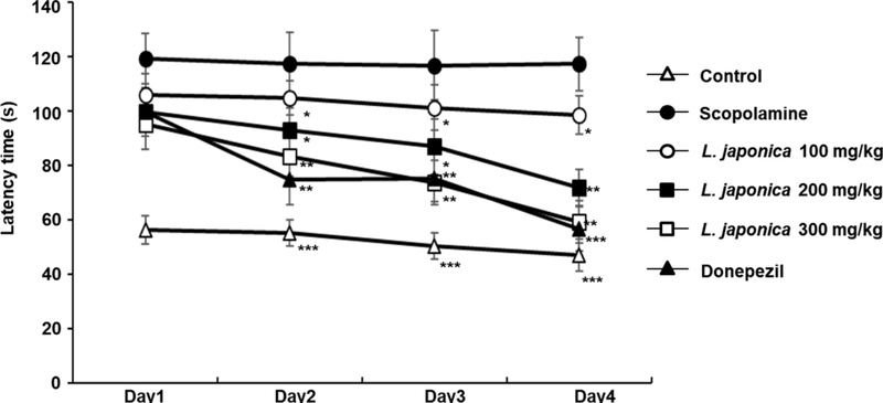
Effects of L. japonica extract on spatial learning and memory in the Morris water maze test. Escape latency was measured in mice with scopolamine-induced memory impairment following oral administration of L. japonica extract (100, 200, and 300 mg/kg) or Donepezil (1 mg/kg), given 90 min prior to scopolamine treatment. The latency to locate the hidden platform during the training sessions is presented. Data are expressed as mean ± SD (n = 6). Statistical significance is indicated as follows: p < 0.05, *p < 0.01, and **p < 0.001 compared with the scopolamine-treated group.
As shown in Fig. 2, clear differences in total swimming distance were observed among the experimental groups, reflecting variations in spatial learning performance and search efficiency during the Morris water maze task. The control group displayed relatively efficient navigation, with a shorter cumulative swimming distance (769 cm), indicating that these animals were able to rapidly learn and remember the location of the hidden platform and adopt a more direct search strategy over repeated trials. In contrast, the scopolamine-treated group exhibited a marked increase in total swimming distance (1192 cm), suggesting severe impairment in spatial learning and memory. These animals showed inefficient and dis-organized swimming patterns, consistent with difficulty in acquiring or retaining the platform location, resulting in increased exploratory behavior and prolonged search paths. Treatment with L. japonica extract significantly reduced the scopolamine-induced increase in swimming distance in a dose-dependent manner across the training sessions. Mice administered 100 mg/kg body weight showed a moderate reduction in swimming distance (1099 cm), while the 200 mg/kg body weight group demonstrated further improvement, with a decrease to 963 cm. The most pronounced effect was observed in the 300 mg/kg body weight group, which exhibited a substantial reduction in swimming distance to 854 cm, approaching a more efficient search pattern compared with the scopolamine group. Overall, these results indicate that L. japonica extract improves spatial learning ability and enhances navigation efficiency, enabling mice to locate the hidden platform with less redundant movement and reduced overall swimming distance (Fig. 2).
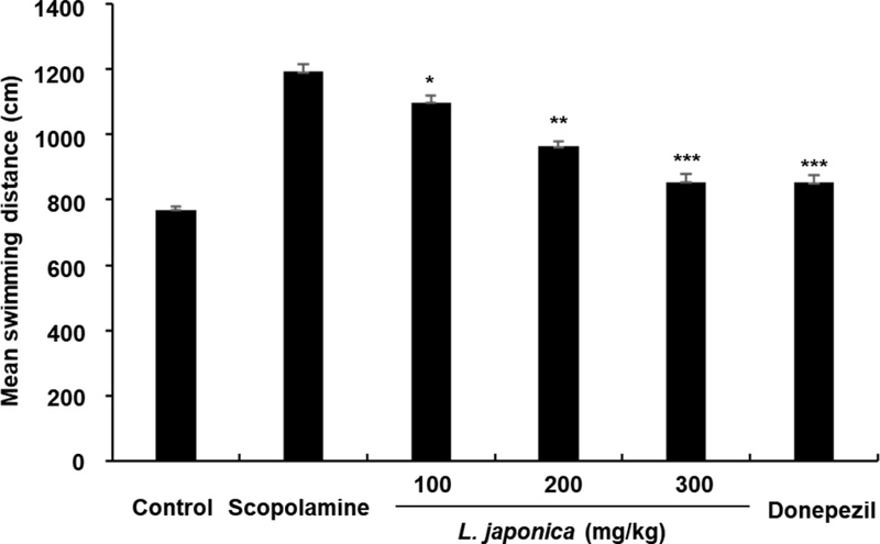
The effect of L. japonica extract on mean swimming distance to find the platform over 4 days. Data are mean escape latencies ± SD (n = 6) (*p < 0.05, **p < 0.01 and ***p < 0.001 versus scopolamine-treated mice).
In the probe trial of the Morris water maze, spatial memory retention was assessed by quantifying the time spent in the target quadrant where the escape platform had previously been located (Fig. 3). The control group showed strong memory retention, spending significantly more time in the target quadrant (29.3 s), indicating successful consolidation of spatial memory and accurate recall of the platform location. In contrast, scopolamine administration resulted in a pronounced impairment of memory retention, as evidenced by a substantial reduction in target quadrant exploration time (10.5 s). This reduction reflects a failure to retain spatial information acquired during the training phase, consistent with disrupted cholinergic signaling and memory dysfunction. Treatment with L. japonica extract significantly ameliorated these deficits in a dose-dependent manner. Mice treated with 100 mg/kg body weight and 200 mg/kg body weight of the extract showed moderate improvements, spending 17.3 s and 20.5 s in the target quadrant, respectively, suggesting partial restoration of memory retention. Notably, the 300 mg/kg body weight group exhibited the most robust effect, with a marked increase to 24.5 s, approaching the level observed in the control group. Overall, these results indicate that L. japonica extract effectively improves scopolamine-induced impairments in spatial memory retention, as demonstrated by enhanced preference for the target quadrant during the probe trial (Fig. 3).
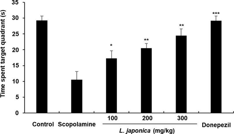
The effect of L. japonica extract in the probe trial. The time spent in the target quadrant during the probe trial was presented. Data are mean escape latencies ± SD (n = 6) (*p < 0.05, **p < 0.01 and ***p < 0.001 versus scopolamine-treated mice).
To rule out the possibility that differences in behavioral performance were due to alterations in locomotor activity, the average swimming speed of mice was analyzed across all experimental groups (Fig. 4). As shown in the results, no statistically significant differences in swimming velocity were observed among the control, scopolamine-treated, donepezil-treated, and L. japonica extract-treated groups. These findings indicate that neither scopolamine administration nor treatment with donepezil or L. japonica extract had a measurable effect on general motor function or swimming ability. All groups exhibited comparable locomotor activity throughout the testing period, suggesting that the animals maintained normal physical performance regardless of treatment. Accordingly, the improvements observed in escape latency, swimming distance, and probe trial performance in the treatment groups cannot be attributed to enhanced motor function or increased swimming speed. Instead, these behavioral changes are more likely to reflect genuine enhancements in cognitive processes, particularly spatial learning and memory, induced by L. japonica extract.
The neuroprotective effect of L. japonica extract against scopolamine-induced cognitive impairment was further evaluated using the Morris water maze (MWM) test, a widely accepted behavioral paradigm for assessing spatial learning and memory performance.23 Scopolamine is known to induce cognitive deficits by antagonizing muscarinic acetylcholine receptors, thereby disrupting cholinergic neurotransmission. In addition, it has been reported to increase acetylcholinesterase (AChE) activity in the hippocampus, which further contributes to reduced acetylcholine availability and impaired memory function.24
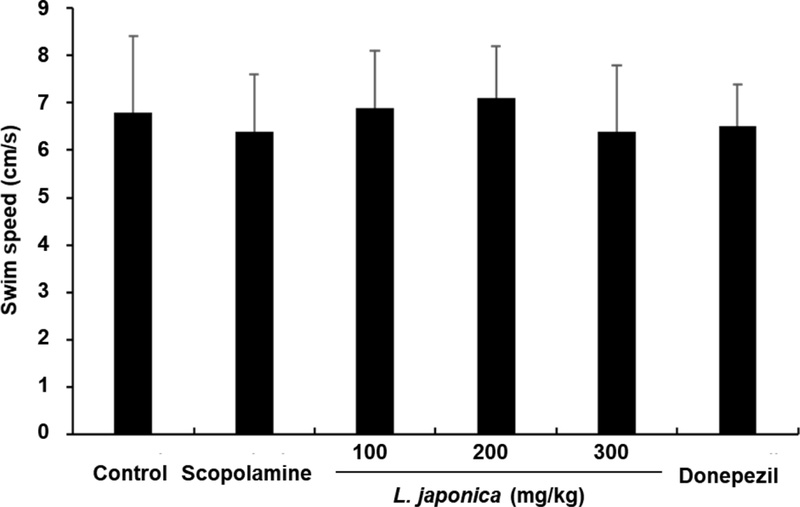
The effect of L. japonica extract on swim speed to find the platform over 4 days. Data are mean escape latencies ± SD (n = 6).
In the present study, treatment with L. japonica extract significantly reduced the scopolamine-induced prolongation of escape latency in a dose-dependent manner. Animals receiving the extract showed progressively improved performance over the training sessions, indicating enhanced acquisition of spatial learning and more efficient navigation toward the hidden platform. These improvements suggest that the extract facilitates learning processes impaired by cholinergic dysfunction.
Importantly, analysis of swimming speed revealed no significant differences among all experimental groups throughout the testing period. This indicates that neither scopolamine nor the treatments influenced general locomotor activity, confirming that the observed behavioral improvements were not attributable to changes in motor function or physical ability. Furthermore, in the probe trial, mice treated with L. japonica extract spent a significantly longer time in the target quadrant compared with the scopolamine group, demonstrating improved memory retention and more accurate recall of the platform location. Collectively, these findings suggest that L. japonica extract effectively alleviates scopolamine-induced deficits in both spatial learning acquisition and memory consolidation.
The passive avoidance test, a well-established behavioral paradigm for assessing associative learning and long-term memory retention, was conducted to further examine the cognitive effects of L. japonica extract in a scopolamine-induced amnesia model (Fig. 5). This test evaluates the ability of animals to remember an aversive stimulus and avoid a previously punished environment.
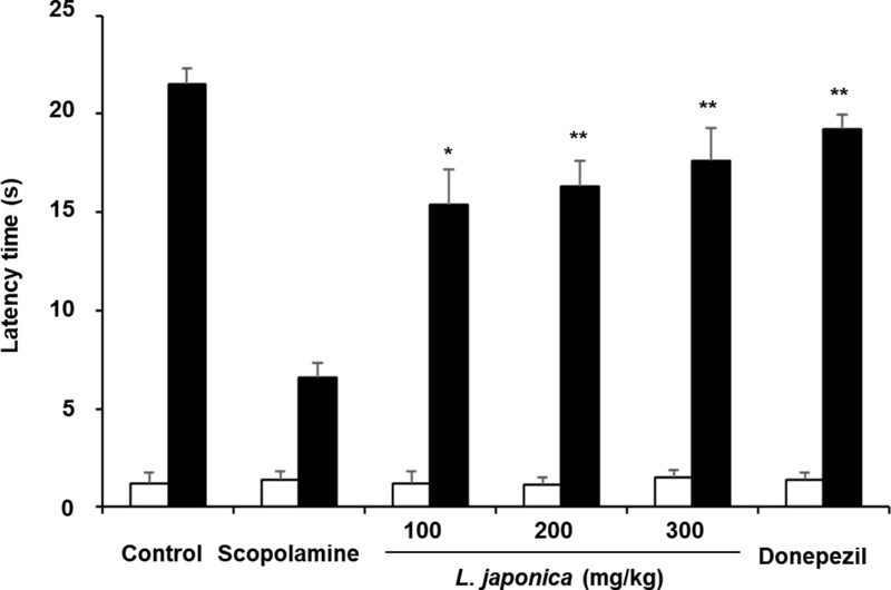
The effect of L. japonica extract on scopolamine-induced memory impairment in the passive avoidance test. The latency prior to entry to the dark compartment was recorded. Data are mean latency times (s) ± SD (n = 6). (*p < 0.05 and **p < 0.01 compared with the scopolamine treated group).
During the acquisition phase, step-through latency did not differ significantly among all experimental groups, indicating comparable baseline exploratory behavior prior to memory formation. The control, scopolamine-treated, and donepezil-treated groups exhibited latency values of 1.2 s, 1.4 s, and 1.4 s, respectively, while the L. japonica extract-treated groups showed similar initial responses with values of 1.2 s, 1.1 s, and 1.5 s at doses of 100, 200, and 300 mg/kg body weight. These results confirm that no pre-existing differences were present among groups before training. In the retention (test) phase, a clear impairment in memory performance was observed in the scopolamine-treated group, as evidenced by a significant reduction in step-through latency compared with the control group. The control animals exhibited a latency of 21.5 s, whereas scopolamine administration markedly decreased this value to 6.6 s, confirming successful induction of cognitive dysfunction. In contrast, treatment with L. japonica extract significantly improved memory retention in a dose-dependent manner. The latency times increased to 15.4 s, 16.3 s, and 17.6 s in the 100, 200, and 300 mg/kg body weight groups, respectively, indicating partial to substantial recovery of associative memory. Although none of the treatment groups fully restored latency to control levels, the highest dose showed the most pronounced improvement. Overall, these findings demonstrate that L. japonica extract effectively attenuates scopolamine-induced impairments in long-term associative memory, as reflected by enhanced avoidance behavior in the passive avoidance test.
The passive avoidance test is a classical behavioral paradigm used to assess associative learning and long-term memory, in which animals learn to avoid an environment previously paired with an aversive stimulus such as a mild electric foot shock. In the present study, L. japonica extract was administered orally to determine its potential to alleviate scopolamine-induced cognitive deficits and improve memory retention. During the acquisition phase, no significant differences in step-through latency were observed among all experimental groups, indicating that baseline exploratory behavior and initial learning conditions were comparable prior to conditioning. This suggests that neither scopolamine nor the administered treatments influenced the animals’ innate activity or initial response to the apparatus. However, in the retention phase, clear group differences were observed. Mice treated with L. japonica extract exhibited a dose-dependent increase in step-through latency compared with the scopolamine-treated group, indicating improved recall of the aversive stimulus and enhanced avoidance behavior. The increase in latency reflects a restoration of memory retention capacity that had been impaired by scopolamine administration. Overall, these findings indicate that L. japonica extract effectively mitigates scopolamine-induced deficits in associative learning and long-term memory, suggesting its potential to enhance cognitive performance under conditions of cholinergic dysfunction.
The effect of L. japonica extract on hippocampal acetylcholinesterase (AChE) activity was further examined to clarify its influence on cholinergic neurotransmission (Fig. 6). As expected, scopolamine administration significantly increased AChE activity in the hippocampus, reaching 165.3% of the control level, indicating a pronounced disruption of cholinergic signaling associated with memory impairment. In contrast, oral administration of L. japonica extract effectively suppressed the scopolamine-induced elevation of AChE activity in a dose-dependent manner (p < 0.05). AChE activity was reduced to 141.2%, 119.5%, and 112.1% of the control level in the 100, 200, and 300 mg/kg treatment groups, respectively. Although enzyme activity remained above the baseline control level, the highest dose exhibited a substantial normalization effect compared with the scopolamine-only group. These results suggest that L. japonica extract may exert its cognitive-enhancing effects, at least in part, through modulation of the cholinergic system via inhibition of AChE activity in the hippocampus, thereby contributing to improved acetylcholine availability and synaptic transmission. To better understand the mechanism underlying the cognitive-enhancing effects of L. japonica extract, both enzymatic activity and cholinergic regulation were examined. Acetylcholine (ACh) is a critical neurotransmitter involved in higher cognitive processes, including learning, memory formation, and particularly spatial and working memory functions. In neurodegenerative disorders such as Alzheimer’s disease, a significant reduction in acetylcholine levels is commonly observed and is strongly correlated with progressive cognitive decline. It has been well established that increased acetylcholinesterase (AChE) activity contributes to the accelerated hydrolysis of acetylcholine, thereby exacerbating cholinergic dysfunction in the brain.25,26 For this reason, AChE has been widely recognized as an important pharmacological target for the treatment of cognitive impairment. Clinically used AChE inhibitors, including donepezil, galantamine, and tacrine, function by preventing acetylcholine breakdown and thereby enhancing cholinergic neurotransmission.26,27
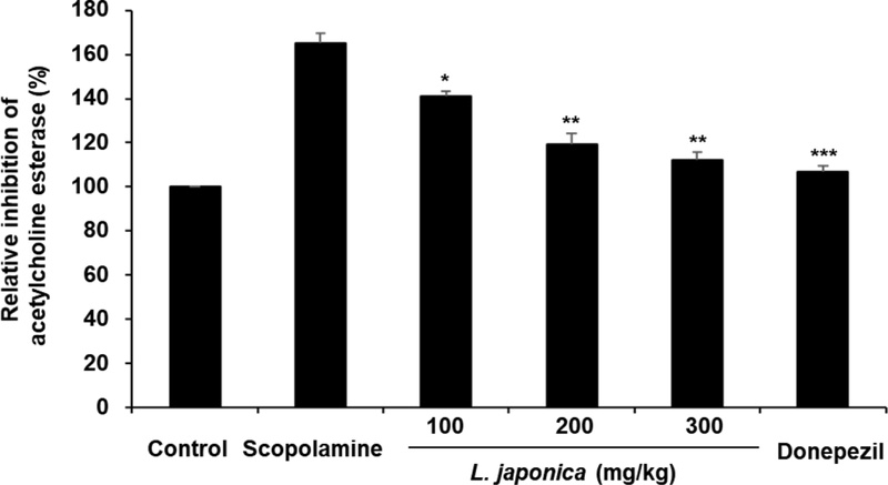
The effect of L. japonica extract on activity of acetylcholine esterase in the hippocampi of the mice. Data were means ± SD. *p < 0.05, **p < 0.01 and ***p < 0.001 compared with the scopolamine-treated group (n = 3).
In the present study, L. japonica extract exhibited a clear dose-dependent inhibitory effect on hippocampal AChE activity. This suppression of enzymatic activity suggests that the extract may help maintain higher levels of acetylcholine within the synaptic cleft, thereby supporting improved cholinergic signaling. Collectively, these findings indicate that the cognitive-enhancing effects of L. japonica extract are likely mediated, at least in part, through modulation of the cholinergic system via inhibition of AChE, leading to enhanced acetylcholine availability in the brain and improved cognitive function under conditions of scopolamine-induced impairment. Recent phytochemical studies on L. japonica have identified a variety of bioactive constituents, including flavonoids such as lonicerin, luteolin, and kaempferol glycosides, as well as phenolic compounds like caffeic acid and chlorogenic acid. These compounds have been reported to exert multiple neuroprotective effects in neuronal systems. In particular, they are known to reduce oxidative stress, regulate intracellular Ca2+ homeostasis, and maintain mitochondrial integrity, all of which are critical factors for neuronal survival and function.28–30
Considering that oxidative stress, mitochondrial dysfunction, and calcium dysregulation are key pathological features involved in neurodegenerative processes, including Alzheimer’s disease, these phytochemicals are likely to play an important role in the cognitive benefits observed in the present study. Among them, flavonoids such as luteolin and kaempferol derivatives have been reported not only to scavenge reactive oxygen species but also to inhibit acetylcholinesterase activity, thereby contributing to enhanced cholinergic neurotransmission and improved learning and memory performance.31–33
Furthermore, phenolic acids including caffeic acid and chlorogenic acid possess strong antioxidant and anti-inflammatory properties. These compounds have also been shown to protect neuronal cells against glutamate-induced excitotoxicity and to ameliorate cognitive dysfunction in experimental models.34–36
Taken together, the cognitive-enhancing effects observed with L. japonica extract in the present study may be attributed to the combined and possibly synergistic actions of these bioactive constituents. These effects are likely mediated through the reinforcement of endogenous antioxidant defense systems, suppression of neuroinflammatory responses, and modulation of the cholinergic system via acetylcholinesterase inhibition, ultimately contributing to improved neuronal function and memory performance.
In conclusion, the present study demonstrates that L. japonica extract effectively attenuates scopolamine-induced cognitive deficits in mice, as evidenced by significant improvements in both spatial and associative learning performance across behavioral paradigms. These beneficial effects appear to be closely linked to the regulation of cholinergic neurotransmission, particularly through the suppression of acetylcholinesterase activity, which may contribute to the restoration of acetylcholine availability in the brain.
Taken together, these findings suggest that L. japonica extract may serve as a promising natural candidate for the prevention and management of neurodegenerative conditions, including Alzheimer’s disease, where cholinergic dysfunction plays a central role in cognitive decline.
However, the exact bioactive constituents responsible for these observed effects have not yet been fully identified. Although several phytochemicals have been proposed, their individual contributions and potential synergistic interactions remain unclear. Therefore, further studies are warranted to isolate, purify, and structurally characterize the active compounds within the extract. In addition, subsequent investigations should include in vivo validation of these isolated constituents to confirm their efficacy, pharmacological mechanisms, and therapeutic relevance in cognitive impairment models.
Acknowledgments
This research was supported by Korea Basice Science Institute (National research Facilities and Equipment Center) grant funded by the Ministry of Education (grant No. 2022R1A6C101A739).
Conflict of Interest Statement
The authors have declared that there are no conflicts of interest.
References
-
Prince, M.; Bryce, R.; Albanese, E.; Wimo, A.; Ribeiro, W.; Ferri, C. P. Alzheimers Dement. 2013, 9, 63–75.
[https://doi.org/10.1016/j.jalz.2012.11.007]

-
Querfurth, H. W.; LaFerla, F. M. N. Engl. J. Med. 2010, 362, 329–344.
[https://doi.org/10.1056/NEJMra0909142]
-
Hardy, J.; Selkoe, D. J. Science 2002, 297, 353–356.
[https://doi.org/10.1126/science.1072994]
-
Wang, Y.; Mandelkow, E. Nat. Rev. Neurosci. 2016, 17, 5–21.
[https://doi.org/10.1038/nrn.2015.1]
- Birks, J. S. Cochrane Database Syst. Rev. 2006, 2006, CD005593.
- Birks, J.; Grimley Evans, J. Cochrane Database Syst. Rev. 2015, 10, CD001191.
- Loy, C.; Schneider, L. Cochrane Database Syst. Rev. 2006, 18, CD001747.
-
Kumar, A.; Singh, A.; Ekavali Pharmacol. Rep. 2015, 67, 195–203.
[https://doi.org/10.1016/j.pharep.2014.09.004]
-
Morris, R. J. Neurosci. Methods 1984, 11, 47–60.
[https://doi.org/10.1016/0165-0270(84)90007-4]
-
Bevins, R. A.; Besheer, J. Nat. Protoc. 2006, 1, 1306–1311.
[https://doi.org/10.1038/nprot.2006.205]
-
Shang, X.; Pan, H.; Li, M.; Miao, X.; Ding, H. J. Ethnopharmacol. 2011, 138, 1–21.
[https://doi.org/10.1016/j.jep.2011.08.016]
- Chen, J.; Li, W.; Yao, H.; Chin. J. Nat. Med. 2015, 13, 321–331.
-
Shang, X.; Pan, H.; Li, M.; Miao, X.; Ding, H. J. Ethnopharmacol. 2011, 138, 1–21.
[https://doi.org/10.1016/j.jep.2011.08.016]
- Liu, A. L.; Liu, B.; Qin, H. L.; Lee, S. M.; Wang, Y. T.; Du, G. H. J. Ethnopharmacol. 2008, 116, 245–249.
- Wang, Z.; Clifford, M. N.; Sharp, P. J. Agric. Food Chem. 2008, 56, 394–399.
- Kang, O. H.; Choi, J. G.; Lee, Y. S.; Kwon, D. Y. Food Chem. Toxicol. 2010, 48, 212–217.
- Li, Y.; But, P. P. H.; Ooi, V. E. C. J. Ethnopharmacol. 2005, 96, 475–480.
- Park, K. I.; Park, H. S.; Nagappan, A. Int. J. Mol. Med. 2012, 29, 1001–1006.
-
Choi, C. W.; Jung, H. A.; Kang, S. S.; Choi, J. S. Arch. Pharm. Res. 2007, 30, 1–7.
[https://doi.org/10.1007/BF02977770]
-
Li, H. B.; Jiang, Y.; Chen, F. Food Chem. 2008, 106, 2–10.
[https://doi.org/10.1016/j.foodchem.2007.04.067]
- Zhang, Y.; Han, T.; Yu, C.; Rahman, K.; Qin, L.; Peng, C. J. Ethnopharmacol. 2016, 189, 1–10.
- Colović, M. B.; Krstić, D. Z.; Lazarević‐Pašti, T. D.; Bondžić, A. M.; Vasić, V. M. Curr. Neuropharmacol. 2013, 11, 315–335.
-
Morris, R. J. Neurosci. Methods. 1984, 11, 47–60.
[https://doi.org/10.1016/0165-0270(84)90007-4]
-
Klinkenberg, I.; Blokland, A. Neurosci. Biobehav. Rev. 2010, 34, 1307–1350.
[https://doi.org/10.1016/j.neubiorev.2010.04.001]
-
Ferreira‐Vieira, T. H.; Guimaraes, I. M.; Silva, F. R.; Ribeiro, F. M. Curr. Neuropharmacol. 2016, 14, 101–115.
[https://doi.org/10.2174/1570159X13666150716165726]
-
Francis, P. T.; Palmer, A. M.; Snape, M.; Wilcock, G. K. J. Neurol. Neurosurg. Psychiatry 1999, 66, 137–147.
[https://doi.org/10.1136/jnnp.66.2.137]
-
Hampel, H.; Mesulam, M.‐M.; Cuello, A. C.; Farlow, M. R.; Giacobini, E.; Grossberg, G. T.; Khachaturian, A. S.; Vergallo, A.; Cavedo, E.; Snyder, P. J.; Khachaturian, Z. S. Brain 2018, 141, 1917–1933.
[https://doi.org/10.1093/brain/awy132]
- Lin, L. M.; Zhang, X. G.; Zhu, J. J.; Gao, H. M.; Wang, Z. M.; Wang, W. H. J. Ethnopharmacol. 2008, 116, 186–190.
-
Lopez‐Lazaro, M. Mini Rev. Med. Chem. 2009, 9, 31–59.
[https://doi.org/10.2174/138955709787001712]
-
Spencer, J. P. E. Proc. Nutr. Soc. 2010, 69, 244–260.
[https://doi.org/10.1017/S0029665110000054]
-
Orhan, I.; Kartal, M.; Tosun, F.; Şener, B. Food Chem. 2007, 104, 630–635.
[https://doi.org/10.1016/j.foodchem.2006.12.012]
- Dajas, F.; Rivera‐Megret, F.; Blasina, F.; Arredondo, F.; Echeverry, C.; Lafon, L.; Heizen, H.; Ferreira, M.; Morquio, A. J. Pharmacol. Exp. Ther. 2003, 304, 713–720.
- Nabavi, S. F.; Braidy, N.; Gortzi, O.; Sobarzo‐Sánchez, E.; Daglia, M.; Skalicka‐Woźniak, K.; Nabavi, S. M. CNS Neurol. Disord. Drug Targets 2015, 14, 860–868.
- Lee, K. J.; Choi, J. H.; Kim, H. G.; Han, E. H.; Jeong, H. G. Food Chem. Toxicol. 2008, 46, 1655–1661.
-
Kono, Y.; Kobayashi, K.; Tagawa, S.; Adachi, K.; Ueda, A.; Sawa, Y.; Shibata, H. Biochim. Biophys. Acta 1997, 1335, 335–342.
[https://doi.org/10.1016/S0304-4165(96)00151-1]
- Oboh, G.; Akinyemi, A. J.; Ademiluyi, A. O. J. Basic Clin. Physiol. Pharmacol. 2012, 23, 79–85.