
Anti-pemphigus and Anti-atopic Potentials of Machilus thunbergii Cortex Extract with MLK3 Inhibitory Activity in Human Keratinocyte HaCaT Cells
Abstract
Pemphigus is an autoimmune blistering disease characterized by autoantibody-mediated disruption of desmosomal adhesion in keratinocytes. Accumulating evidence suggests that intracellular signaling pathways, including mixed lineage kinase 3 (MLK3)–mediated MAPK activation, play a critical role in pemphigus pathogenesis. In this study, we investigated the effects of Machilus thunbergii cortex extract (EMTC) on MLK3 signaling and keratinocyte dysfunction in experimental models of pemphigus. Treatment of HaCaT keratinocytes with the pathogenic anti-desmoglein 3 antibody AK23 induced rapid, time-dependent phosphorylation of MLK3. EMTC significantly inhibited MLK3 activity in a dose-dependent manner without affecting cell viability. Moreover, EMTC attenuated AK23-induced cellular fragmentation, indicating a protective effect against keratinocyte adhesion loss. In addition to its effects on desmosomal destabilization, EMTC suppressed TNF-α/IFN-γ–induced production of thymus and activation-regulated chemokine (TARC), suggesting an anti-inflammatory role in keratinocytes. Collectively, these findings demonstrate that EMTC mitigates pemphigus-associated keratinocyte damage by targeting MLK3-mediated signaling pathways and inflammatory responses. Our results suggest that Machilus thunbergii cortex extract may represent a novel, pathway-targeted therapeutic candidate for the treatment of pemphigus.
Keywords:
Pemphigus, MLK3, MAPK signaling, Machilus thunbergii cortex extract (EMTC), KeratinocytesIntroduction
Pemphigus is a rare autoimmune blistering disease characterized by the formation of intraepidermal blisters and erosions affecting the skin and mucous membranes. The disease is primarily caused by pathogenic autoantibodies directed against desmosomal cadherins such as desmoglein 3 (Dsg3), leading to impaired keratinocyte cell–cell adhesion and subsequent acantholysis.1–4 Although the role of autoantibodies in pemphigus has been well established, the intracellular signaling mechanisms that mediate desmosome disassembly and blister formation remain incompletely understood.
Recent studies have demonstrated that intracellular signaling cascades, particularly mitogen-activated protein kinase (MAPK) pathways, play a critical role in the pathogenesis of pemphigus.5–7 Activation of MAPK family members, including p38 MAPK, ERK, and Src, has been reported in keratinocytes exposed to pemphigus autoantibodies. Pharmacological inhibition of these pathways significantly attenuates blister formation in experimental models, highlighting their importance in disease progression and identifying them as potential therapeutic targets.
Mixed lineage kinase 3 (MLK3), also known as mitogen-activated protein kinase kinase kinase 11 (MAP3K11), is a serine/threonine kinase that functions as an upstream regulator of multiple MAPK pathways, including JNK, p38 MAPK, and ERK. MLK3 has been extensively studied in cancer biology, where it promotes tumor cell survival, invasion, and metastasis through activation of stress- and inflammation-related signaling pathways.8–10 Beyond oncology, recent studies have suggested that MLK3-mediated signaling may contribute to epithelial barrier dysfunction by disrupting cell–cell adhesion.11,12 In pemphigus, activation of MLK3 has been shown to induce phosphorylation of downstream effectors such as ERK and c-Jun, ultimately leading to desmosomal destabilization and loss of intercellular adhesion.13,14
Considering the shared involvement of MLK3 and MAPK signaling in cancer progression and autoimmune blistering diseases, targeting these pathways may provide a novel and effective therapeutic strategy. However, the development of safe and effective inhibitors remains challenging, prompting interest in bioactive natural compounds with pathway-modulating properties.
Machilus thunbergii (Lauraceae), commonly known as Tohubak in Korea, is an evergreen tree widely distributed in East Asia, including Korea and Japan. The stem bark of M. thunbergii has been traditionally used in Korean herbal medicine for the treatment of gastrointestinal disorders, inflammation, and pain.15,16
Phytochemical studies have reported that M. thunbergii contains various bioactive constituents, including lignans, neolignans, alkaloids, and flavonoids, which exhibit anti-inflammatory, antioxidant, and antimicrobial activities. Consistent with these findings, recent pharmacological studies have demonstrated that crude extracts and solvent fractions of M. thunbergii possess diverse biological activities, such as anti-inflammatory, antioxidant, neuroprotective, and hepatoprotective effects.17,18 In particular, in lipopolysaccharide-stimulated macrophages, M. thunbergii extract suppresses the production of inflammatory mediators, including nitric oxide, prostaglandin E₂, tumor necrosis factor-α, and interleukin-6, through inhibition of NF-κB and p38 MAPK signaling pathways.19 Furthermore, extracts derived from the bark have been reported to protect cells against oxidative stress and inflammatory responses, suggesting their potential therapeutic applications in inflammatory and immune-related diseases.
Despite the growing pharmacological evidence supporting the therapeutic potential of M. thunbergii, its effects on pemphigus-associated signaling pathways, particularly MLK3 and MAPK cascades, have not yet been fully explored. In the current study, we investigated the effects of Machilus thunbergii cortex extract (EMTC) on MLK3-mediated MAPK signaling and keratinocyte cell–cell adhesion in experimental models of pemphigus, to explore its potential relevance to therapeutic modulation of this autoimmune skin disease.
Experimental
Preparation of Machilus thunbergii cortex extract – The plant extract (CA04-069) used in this study was supplied by the Korea Plant Extract Bank, Korea Research Institute of Bioscience and Biotechnology (KRIBB; Daejeon, Republic of Korea). A voucher specimen (PBC-459) has been deposited in the herbarium of KRIBB. Shade-dried plant material (110 g) was finely ground and extracted with 1 L of 95.0% ethyl alcohol (GR grade). Extraction was carried out at room temperature using an ultrasonic extractor (SDN-900H, SD-ULTRASONIC CO., LTD) under the following conditions: 30 extraction cycles consisting of 15 min of ultrasonication (40 kHz, 1500W) followed by 120 min of standing per cycle. The extract was subsequently filtered using qualitative filter paper (No.100, HYUNDAI MICRO CO., LTD) and concentrated under reduced pressure. This procedure yielded 8.56 g of M. thunbergii cortex extract.
Cell culture – Immortalized human keratinocyte HaCaT cells (AddexBio, CA, USA) were maintained in low-glucose DMEM supplemented with 10% fetal bovine serum and 1% antibiotic-antimycotic solution under standard culture conditions (37°C, 5% CO₂). All reagents used for cell culture were purchased from HyClone (UT, USA).
Western blot analysis – HaCaT cells were plated at a density of 5 × 10⁵ cells per well in 6-well plates and allowed to adhere for 24 h. Cells were then exposed to 5 μg/mL of the anti-desmoglein 3 monoclonal antibody AK23 (IgG clone; MBL, Japan) for the indicated durations. After treatment, cells were rinsed with PBS and lysed on ice using RIPA lysis buffer (ELPIS Biotech, Korea) supplemented with protease and phosphatase inhibitor cocktails (Thermo Fisher Scientific, MA, USA). Cell lysates were clarified by centrifugation at 13,000 rpm for 30 min at 4°C, and protein concentrations in the supernatants were determined using the Pierce BCA Protein Assay Kit (Thermo Fisher Scientific). Equal amounts of protein (10 μg) were separated by SDS-PAGE using 5–20% gradient gels (ATTO, Tokyo, Japan) at 130 V for 90 min and subsequently transferred onto polyvinylidene difluoride membranes (ATTO). Membranes were blocked with TBST containing 5% bovine serum albumin for 1 h at room temperature, followed by incubation with primary antibodies overnight at 4°C. All primary antibodies were obtained from Cell Signaling Technology (MA, USA), and GAPDH was used as a loading control. After washing three times with TBST, membranes were incubated with HRP-conjugated secondary antibodies for 2 h at room temperature. Immunoreactive signals were visualized using enhanced chemiluminescence reagents (Millipore, MA, USA) and detected with a WSE-6100 LuminoGraph imaging system (ATTO).
MLK3 kinase activity assay – MLK3 kinase activity assays were performed by Eurofins (France). Recombinant human MLK3 was reacted in a buffer containing 8 mM MOPS pH 7.0, 0.2 mM EDTA, 0.33 mg/mL myelin basic protein as the MLK3 subtrate, 5 mM DTT, 10 mM Magnesium acetate, together with 45 mM [γ-33P]ATP. The enzymatic reaction was initiated by adding the Mg/ATP mixture and allowed to proceed for 120 min at room temperature. The reaction was terminated by the addition of phosphoric acid to a final concentration of 0.5%. Portions of the reaction mixture were then applied to filter membranes, followed by four washes (4 min each) with 0.425% phosphoric acid and a final wash with methanol. After drying, incorporated radioactivity was quantified by scintillation counting.
Cell viability assay – HaCaT keratinocytes were seeded into 96-well plates (4 × 10³ cells/well) and cultured for 24 h prior to treatment. The cells were then treated with EMTC for 24 h, after which cell viability was evaluated using the CCK-8 assay (LPSolution). Measurements were performed in triplicate, and absorbance at 450 nm was measured using a HIDEX Sense plate reader. Cell viability data were used to generate dose-response curves with GraphPad Prism software (version 5).
Dispase-based fragmentation assay – For dissociation analysis, HaCaT cells were plated in 24-well plates (5 × 10⁵ cells/well) and allowed to reach confluence over 24 h. Cells were exposed to AK23 (5 μg/mL) either alone or in combination with EMTC for 24 h. Following treatment, monolayers were gently washed with warm PBS and incubated with Dispase II (2.4 U/mL) diluted in HBSS for 20 min at 37°C. Mechanical disruption was applied by repeated pipetting (15 strokes) after the addition of fresh HBSS. Dissociated cell fragments were subsequently stained with MTT and counted, and images were captured using microscopy. Experiments were performed in triplicate, and data were analyzed using Student’s t-test, with statistical significance defined as p < 0.05.
Enzyme-linked immunosorbent assay – HaCaT keratinocytes (1 × 10⁶ cells/well) were seeded into 6-well plates and maintained in DMEM containing 10% fetal bovine serum. Cells were pretreated with EMTC for 1 h prior to co-stimulation with TNF-α and IFN-γ (each at 5 ng/mL) for 24 h. The concentration of TARC (CCL17) released into the culture medium was determined by sandwich ELISA using a commercially available human CCL17/TARC Quantikine kit (R&D Systems) following the supplier’s instructions.
Results and Discussion
To investigate the effect of AK23 on MLK3 activation, HaCaT cells were treated with AK23 (5 μg/mL) for the indicated time points and analyzed by western blotting. As shown in Fig. 1, phosphorylated MLK3 (p-MLK3) levels were markedly increased at 30 min after AK23 treatment, whereas no noticeable change was observed at 5 or 15 min compared with the untreated control. In contrast, the expression level of total MLK3 remained unchanged throughout the time course. The loading control, GAPDH, showed comparable expression across all samples, confirming equal protein loading. These results indicate that AK23 induces time-dependent phosphorylation of MLK3 without affecting total MLK3 protein levels.
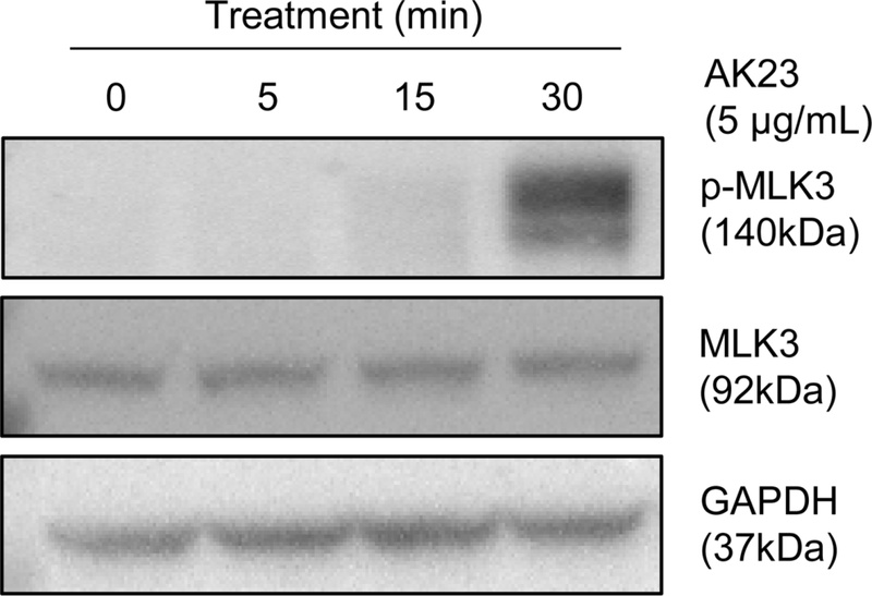
AK23 induces time-dependent phosphorylation of MLK3 in HaCaT cells. HaCaT cells were treated with AK23 (5 μg/mL) for the indicated times (0, 5, 15, and 30 min). Cell lysates were subjected to western blot analysis using antibodies against p-MLK3 and total MLK3. GAPDH was used as a loading control (n = 3).
A library of herbal extracts was screened, and EMTC was identified as an inhibitor of MLK3 kinase activity (data not shown). To directly assess its inhibitory effect on MLK3, kinase activity was measured using recombinant MLK3 protein in a cell-free assay system. As shown in Fig. 2, EMTC inhibited MLK3 activity in a concentration-dependent manner, resulting in a progressive decrease in MLK3 activity with increasing EMTC concentrations. Nonlinear regression analysis revealed an IC50 value of 163 ng/mL, indicating a potent inhibitory effect of EMTC on MLK3 activity.
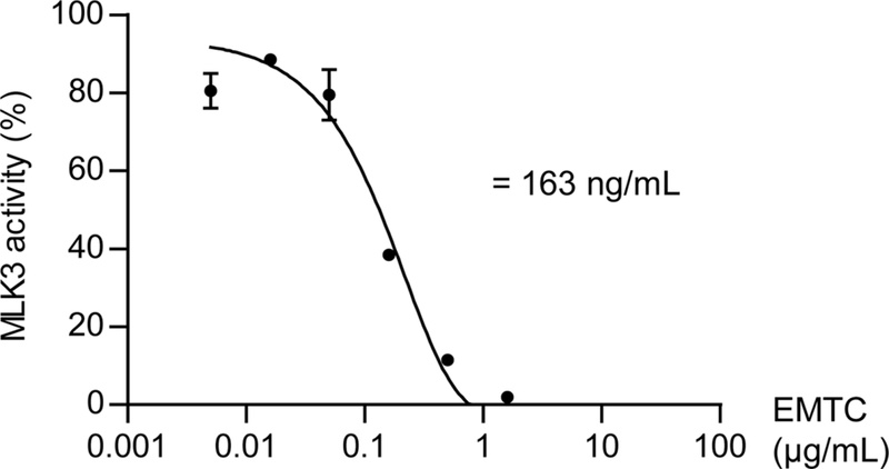
EMTC inhibits MLK3 activity in a dose-dependent manner. MLK3 activity was assessed in the presence of increasing concentrations of EMTC. Data are presented as percentage of MLK3 activity relative to the untreated control. The IC50 value was calculated by nonlinear regression analysis (n = 3).
Prior to assessing the anti-pemphigus potential of EMTC in HaCaT cells, its cytotoxic profile was first examined to rule out the possibility that EMTC-induced cell detachment might result from cellular toxicity rather than specific biological effects. HaCaT cells were exposed to increasing concentrations of EMTC for 24 h, after which cell viability was evaluated using the CCK-8 assay. No significant cytotoxic effects were observed at concentrations up to 30 μg/mL (Fig. 3). Based on these findings, subsequent analyses of the effects of EMTC on pemphigus were conducted at concentrations ≤ 30 μg/mL using a dispase-based fragmentation assay, a well-established in vitro model of pemphigus employing confluent keratinocyte monolayers such as HaCaT cells. The cells were treated with AK23 (5 μg/mL) in the presence or absence of increasing concentrations of EMTC. As shown in Fig. 4, AK23 treatment significantly increased the number of fragments compared with the untreated control. In contrast, co-treatment with EMTC markedly reduced AK23-induced fragmentation in a concentration-dependent manner, with significant decreases observed at 10 and 30 μg/mL. Representative microscopic images further support the quantitative analysis, demonstrating a clear reduction in cellular fragmentation following EMTC treatment. These results indicate that EMTC effectively suppresses AK23-induced cellular fragmentation.
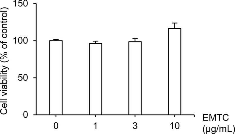
Effect of EMTC on cell viability. Cells were treated with Machilus thunbergii cortex extract (EMTC) at the indicated concentrations (0, 1, 3, and 10 μg/mL). Cell viability was assessed after treatment and expressed as a percentage of the untreated control. Data are presented as mean ± SD (n = 3).
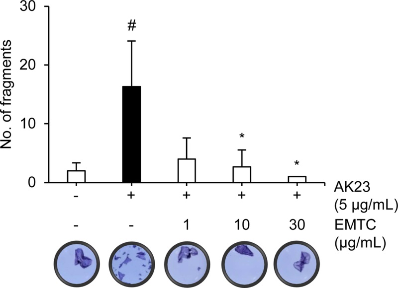
EMTC attenuates AK23-induced cellular fragmentation. Cells were treated with AK23 (5 μg/mL) in the presence of the indicated concentrations of EMTC. The number of cellular fragments was quantified. Data are presented as mean ± SD. Representative images are shown below the graph. Statistical significance: #p < 0.05 versus untreated control; *p < 0.05 versus AK23-treated group (n = 3).
To examine the effect of EMTC on inflammatory chemokine production, cells were stimulated with TNF-α and IFN-γ (5 ng/mL each) in the presence of increasing concentrations of EMTC. As shown in Fig. 5, co-stimulation with TNF-α and IFN-γ markedly increased TARC production compared with the untreated control. In contrast, EMTC treatment significantly reduced TNF-α/IFN-γ-induced TARC levels in a concentration-dependent manner, with significant inhibition observed at concentrations ranging from 1 to 30 μg/mL. These findings indicate that EMTC effectively suppresses inflammatory chemokine production induced by pro-inflammatory cytokines.
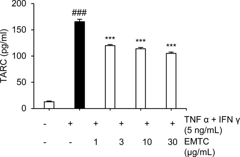
EMTC inhibits TNF-α/IFN-γ-induced TARC production. Cells were stimulated with TNF-α and IFN-γ (5 ng/mL each) in the presence of the indicated concentrations of EMTC. TARC levels in the culture supernatants were measured and expressed as mean ± SD. ###p < 0.001 versus untreated control; ***p < 0.001 versus TNF-α/IFN-γ-treated group (n = 3).
Pemphigus is characterized by autoantibody-mediated disruption of desmosomal adhesion, in which intracellular signaling events play a pivotal role in translating antibody binding into keratinocyte dissociation and blister formation. Although MAPK signaling pathways have been extensively implicated in pemphigus pathogenesis, the upstream regulatory mechanisms governing their activation remain incompletely defined. MLK3 (mixed lineage kinase 3), a member of the MAP3K family, functions as a critical upstream regulator of stress-activated signaling pathways, particularly the JNK and p38 MAPK cascades. MLK3 activation is known to be controlled by multiple upstream stimuli, including small GTPases such as Rac1 and Cdc42, oxidative stress, and TNF-α receptor signaling, all of which are closely associated with inflammatory responses and cytoskeletal reorganization in keratinocytes.20−22 Activation of these pathways promotes JNK/p38 signaling, which has been implicated in keratinocyte detachment and blister formation in pemphigus.23
Recent studies have highlighted MLK3 as an important upstream kinase regulating stress-activated signaling pathways, including the JNK and p38 MAPK cascades, which are involved in inflammatory responses and cytoskeletal remodeling.24,25 Dysregulation of these signaling pathways has been implicated in various pathological conditions, including neurodegeneration, cancer, and inflammatory diseases.25 In the context of pemphigus, activation of MAPK signaling contributes to keratinocyte detachment and acantholysis, suggesting that modulation of upstream kinases such as MLK3 may represent a potential therapeutic strategy. Although several synthetic kinase inhibitors targeting MAPK signaling have been developed, their clinical application is often limited by toxicity and insufficient selectivity due to the highly conserved nature of kinase domains.26
In the present study, we demonstrate that MLK3 is rapidly activated in keratinocytes upon exposure to the pathogenic anti-Dsg3 antibody AK23 and that pharmacological modulation of MLK3 activity by Machilus thunbergii cortex extract (EMTC) effectively attenuates downstream signaling and cellular fragmentation. Our results show that AK23 induces time-dependent phosphorylation of MLK3 without altering total MLK3 protein levels, indicating that MLK3 activation occurs through post-translational regulation rather than changes in expression. This finding is consistent with previous reports implicating MAP3Ks as early signaling hubs in pemphigus-associated signal transduction. Given that MLK3 functions as an upstream activator of multiple MAPK pathways, including ERK, JNK, and p38 MAPK, its rapid activation may represent a critical convergence point linking autoantibody binding to downstream cytoskeletal reorganization and desmosomal destabilization.
Importantly, EMTC directly inhibited MLK3 kinase activity in a cell-free system, with an IC₅₀ value in the nanogram-per-milliliter range, suggesting a potent and specific inhibitory effect. This biochemical inhibition was accompanied by functional protection against AK23-induced keratinocyte fragmentation in HaCaT cells. The ability of EMTC to suppress cellular dissociation supports the notion that MLK3 activity is functionally required for pemphigus-associated loss of intercellular adhesion and highlights MLK3 as a pharmacologically tractable target.
In this regard, natural products have attracted increasing attention as potential modulators of kinase signaling pathways. Plant-derived compounds often exert biological effects through multi-target mechanisms and may regulate upstream signaling networks rather than acting as single-target inhibitors.27 Such characteristics may be particularly beneficial for diseases involving complex signaling cascades, such as autoimmune and inflammatory disorders. Therefore, the identification of MLK3-modulating activity in M. thunbergii extract suggests that natural products may represent a valuable source of novel regulators of MLK3-related signaling pathways. These findings provide a basis for further studies aimed at identifying active constituents and exploring natural product-derived modulators of MLK3 as potential therapeutic candidates.
Notably, phytochemical studies have shown that M. thunbergii contains various lignan compounds, including machilin A, licarin A, meso-dihydroguaiaretic acid, and nectandrin derivatives. Several of these lignans possess strong antioxidant and anti-inflammatory activities. For example, meso-dihydroguaiaretic acid and related lignans have been reported to reduce oxidative stress and lipid peroxidation in cellular and hepatic injury models.28 Because reactive oxygen species (ROS) are known activators of MLK3 signaling, the antioxidant properties of these compounds may suppress MLK3 activation indirectly. In addition, phenolic lignans such as licarin A have been reported to modulate inflammatory signaling pathways, including MAPK and NF-κB pathways. Extracts of M. thunbergii also inhibit the production of inflammatory mediators such as TNF-α and nitric oxide in activated macrophages.19 Since TNF-α signaling represents another upstream trigger of MLK3 activation, suppression of inflammatory cytokine signaling may further contribute to the inhibition of MLK3 expression. Taken together, the lignan-rich composition of M. thunbergii suggests that its components may inhibit MLK3 signaling through modulation of upstream pathways such as oxidative stress and inflammatory signaling.
In addition to its effects on pemphigus-related signaling, EMTC significantly reduced TNF-α/IFN-γ–induced production of the inflammatory chemokine TARC (CCL17) in keratinocytes. Because inflammatory cytokines and chemokines are increasingly recognized as contributors to disease amplification and chronicity in autoimmune skin disorders, the dual effects of EMTC on both adhesion-related signaling and inflammatory mediator production suggest broader immunomodulatory properties. These findings are in line with previous studies reporting anti-inflammatory activities of M. thunbergii-derived compounds and further extend their relevance to keratinocyte-driven skin inflammation.
Taken together, our data support a model in which MLK3 acts as a key upstream regulator of MAPK signaling in pemphigus, mediating autoantibody-induced desmosomal destabilization and keratinocyte fragmentation. By inhibiting MLK3 activity, EMTC effectively attenuates both pathogenic signaling and functional outcomes in experimental models. While further studies are required to identify the active constituents responsible for MLK3 inhibition and to validate these effects in in vivo models, the present findings provide mechanistic insight into the role of MLK3 in pemphigus and suggest that natural product-based modulation of MLK3–MAPK signaling may represent a promising complementary therapeutic approach for autoimmune blistering diseases.
Although the present study demonstrates the anti-pemphigus activity of M. thunbergii extract and its association with MLK3 suppression, several limitations should be considered. Because the experiments were conducted using crude extracts, the specific bioactive constituents responsible for the observed effects remain unclear. Plant extracts typically contain complex mixtures of phytochemicals, and multiple components may contribute synergistically to the biological activity. Therefore, the MLK3 inhibitory effect observed in this study cannot be attributed to a single compound at this stage. Future studies should focus on bioactivity-guided fractionation of the extract to identify the active constituents responsible for MLK3 suppression. In addition, validation of MLK3 inhibitory activity using isolated compounds and kinase-based assays will be necessary to clarify the precise molecular mechanism. Such studies will help determine the key pharmacologically active components of M. thunbergii and further support its therapeutic potential for pemphigus.
Acknowledgments
This research was supported by Korea Research Institute of Chemical Technology (KK2031-10, SI2131-10, SI2231-10, KK2331-10, KK2431-10, KK2531-10 and KK2632-10).
Conflicts of Interest
The authors declare that they have no conflicts of interest.
References
-
Malik, A. M.; Tupchong, S.; Huang, S.; Are, A.; Hsu, S.; Motaparthi, K. Medicina 2021, 57, 1080.
[https://doi.org/10.3390/medicina57101080]

-
Ohata, C.; Ishii, N.; Furumura, M. J. Cutan. Pathol. 2014, 41, 880–889.
[https://doi.org/10.1111/cup.12384]
-
Furue, M.; Kadono, T. Australas. J. Dermatol. 2017, 58, 171–173.
[https://doi.org/10.1111/ajd.12562]
-
Mascaro Jr, J. M.; España, A.; Liu, Z.; Ding, X.; Swartz, S. J.; Fairley, J. A.; Diaz, L. A. Clin. Immunol. Immunopathol. 1997, 85, 90–96.
[https://doi.org/10.1006/clin.1997.4408]
-
Kim, R.; Kim, E. H.; Ahn, M. J.; Choi, Y. W.; Choi, H. Y.; Kim, S. H.; Byun, J. Y. Arch. Dermatol. Res. 2024, 316, 352.
[https://doi.org/10.1007/s00403-024-03114-w]
-
Fuchs, M.; Möchel, M.; Radeva, M. Y.; Schmitt, T., Yazdi, A. S.; Hashimoto, T.; Waschke, J. Br. J. Dermatol. 2025, 193, 468–479.
[https://doi.org/10.1093/bjd/ljaf142]
-
Mavropoulos, A.; Orfanidou, T.; Liaskos, C.; Smyk, D. S.; Spyrou, V.; Sakkas, L. I.; Rigopoulou, E. I.; Bogdanos, D. P. Autoimmune Dis. 2013, 2013, 728529.
[https://doi.org/10.1155/2013/728529]
-
Chen, J.; Gallo, K. A. Cancer Res. 2012, 72, 4130–4140.
[https://doi.org/10.1158/0008-5472.CAN-12-0655]
-
Zhan, Y.; Abi Saab, W. F. A.; Modi, N.; Stewart, A. M.; Liu, J.; Chadee, D. N. Exp. Cell Res. 2012, 318, 1641–1648.
[https://doi.org/10.1016/j.yexcr.2012.05.002]
-
Rattanasinchai, C.; Gallo, K. A. Cancers 2016, 8, 51.
[https://doi.org/10.3390/cancers8050051]
-
Kovalenko, P. L.; Kunovska, L.; Chen, J.; Gallo, K. A.; Basson, M. D. Am. J. Physiol. Gastrointest. Liver Physiol. 2012, 303, G951–G960.
[https://doi.org/10.1152/ajpgi.00158.2012]
-
Rozier, M. C. J.; Adjei, D. N.; Radtke, R. A.; Chadee, D. N. Biochim. Biophys. Acta Mol. Cell Res. 2026, 1873, 120106.
[https://doi.org/10.1016/j.bbamcr.2026.120106]
-
Walter, E.; Vielmuth, F.; Rotkopf, L.; Sárdy, M.; Horváth, O. N.; Goebeler, M.; Schmidt, E.; Eming, R.; Hertl, M.; Spindler, V.; Waschke, J. Sci. Rep. 2017, 7, 3579.
[https://doi.org/10.1038/s41598-017-03697-7]
-
Egu, D. T.; Kugelmann, D.; Waschke, J. Front. Immunol. 2019, 10, 2883.
[https://doi.org/10.3389/fimmu.2019.02883]
-
Kim, S. J.; You, J.; Choi, H. G.; Kim, J. A.; Jee, J.-G.; Lee, S. Phytomedicine 2015, 22, 615–620.
[https://doi.org/10.1016/j.phymed.2015.03.018]
- Lee, K.; P. G.-Y.; Liu, K.-H.; Kim, T.-W.; Ham, I.; Bu, Y.; Choi, H.-Y. Kor. J. Herbology 2011, 26, 75–81.
-
Shin, H.; Han, Y. K.; Byun, Y.; Jeon, Y. H.; Lee, K. Y. Molecules 2021, 26, 4804.
[https://doi.org/10.3390/molecules26164804]
-
Li, G.; Lee, C.-S.; Woo, M.-H.; Lee, S.-H.; Chang, H.-W.; Son, J.-K. Biol. Pharm. Bull. 2004, 27, 1147–1150.
[https://doi.org/10.1248/bpb.27.1147]
-
Wu, S.-J.; Len, W.-B.; Huang, C.-Y.; Liou, C.-J.; Huang, W.-C. Turk. J. Biol. 2015, 39, 657–665.
[https://doi.org/10.3906/biy-1405-50]
-
Teramoto, H.; Coso, O. A.; Miyata, H.; Igishi, T.; Miki, T.; Gutkind, J. S. J Biol Chem. 1996, 271, 27225–27228.
[https://doi.org/10.1074/jbc.271.44.27225]
-
Sathyanarayana, P.; Barthwal, M. K.; Kundu, C. N.; Lane, M. E.; Bergmann, A.; Tzivion, G.; Rana, A. Mol. Cell. 2002, 10, 1527–1533.
[https://doi.org/10.1016/S1097-2765(02)00734-7]
-
Wang, X.; Martindale, J. L.; Liu, Y.; Holbrook, N. J. Biochem. J. 1998, 333, 291–300.
[https://doi.org/10.1042/bj3330291]
-
Ogawa, Y.; Tsuji, M.; Tanaka, E.; Miyazato, M.; Hino, J. Front. Neurol. 2018, 9, 397.
[https://doi.org/10.3389/fneur.2018.00397]
-
Gallo, K. A.; Johnson, G. L. Nat. Rev. Mol. Cell Biol. 2002, 3, 663–672.
[https://doi.org/10.1038/nrm906]
-
Brancho, D.; Ventura, J.-J.; Jaeschke, A.; Doran, B.; Flavell, R. A.; Davis, R. J. Mol. Cell Biol. 2005, 25, 3670–3681.
[https://doi.org/10.1128/MCB.25.9.3670-3681.2005]
-
Collins, C. J.; McCauliff, L. A.; Hyun, S.-H.; Zhang, Z.; Paul, L. N.; Kulkarni, A.; Zick, K.; Wirth, M.; Storch, J.; Thompson, D. H. Biochemistry 2013, 52, 3242–3253.
[https://doi.org/10.1021/bi3010889]
-
Harvey, A. L.; Edrada-Ebel, R.; Quinn, R. J. Nat. Rev. Drug Discov. 2015, 14, 111–129.
[https://doi.org/10.1038/nrd4510]
-
Ma, C. J.; Kim, S. R.; Kim, J.; Kim, Y. C. Br. J. Pharmacol. 2005, 146, 752–759.
[https://doi.org/10.1038/sj.bjp.0706380]