
Assessment of Antioxidant and Neuroprotective Effects of Phyllanthus emblica Extract in a Reserpine-Induced Oxidative Stress Model
Abstract
Amla (Phyllanthus emblica), a nutrient-rich medicinal plant, is renowned for its high antioxidant content, therapeutic properties, and traditional use in Ayurveda to promote health and combat various ailments. This study aimed to determine its total phenol content, antioxidant activity, and mitigating impacts on oxidative stress in an animal model. Total phenol content and antioxidant activity were evaluated using the Folin-Ciocalteu and 2,2-diphenyl-1-picrylhydrazyl (DPPH) methods, respectively. The in vivo study involved 30 female Swiss albino mice subjected to reserpine-induced oxidative stress and treated with varying doses of amla extract (1, 1.5, and 2 mL/kg body weight), with clomipramine as a positive control. In vivo results demonstrated dose-dependent improvements in behavioral (Forced swimming test and tail suspension test immobility time), biochemical (Malondialdehyde and blood glucose), and morphological (brain and adrenal gland weight) parameters. Notably, amla extract significantly attenuated reserpine-induced behavioral despair, reduced lipid peroxidation, and restored brain weight in a dose-dependent manner, indicating marked neuroprotective effects. These findings highlight Bangladeshi-grown amla as a potent natural antioxidant with significant neuroprotective potential in chemically induced oxidative stress, supporting its traditional medicinal use and its possible role in stress-related neurological disorders.
Keywords:
Phyllanthus emblica, Total phenol content, Antioxidants, Oxidative stress, DPPH, Animal modelIntroduction
Oxidative stress, a consequence of an imbalance between reactive oxygen species (ROS) production and the antioxidant defense system, is a major contributing factor to various chronic diseases, including neurodegenerative disorders, cardiovascular diseases, and metabolic syndromes. The quest for natural antioxidants with potent free radical-scavenging abilities has intensified in recent years, driven by the need for safer and more effective therapeutic interventions. Natural antioxidants, particularly those derived from plant sources, are increasingly favored due to their biocompatibility, low toxicity, and broad-spectrum biological activities.1 Fruits (berries, amla, oranges, grapes), vegetables (leafy greens, tomatoes, carrots), nuts, seeds, grains, drinks (green tea, coffee), and spices (coriander, ginger, oregano) are foods high in natural antioxidants.2
Phenolic compounds have emerged as key contributors to antioxidant defense, owing to their ability to donate hydrogen atoms or electrons and chelate metal ions. These can be of many different kinds, including flavonoids (found in fruits, vegetables, tea, and wine), phenolic acids (found in coffee, tea, and berries), tannins (found in tea, wine, and grapes), stilbenes (found in grapes and wine, similar to resveratrol), and lignans (found in seeds, grains, and vegetables ).3 By reducing lipid peroxidation, binding to metal ions that generate free radicals, neutralizing free radicals, and increasing antioxidant enzyme activity, these substances aid in the reduction of oxidative stress.4 Given the tight relationship between oxidative stress and inflammation, phenolic substances also have an anti-inflammatory effect.5
Amla, also known as Indian gooseberry (Phyllanthus emblica), is a widely available fruit in Bangladesh. It is commonly cultivated in various regions due to its adaptability to different climatic conditions. Amla is an integral part of traditional medicine, particularly in Ayurveda, and is widely consumed in different forms, including fresh fruit, juice, dried powder, and pickles. In Bangladesh, it is also used as an integral part of herbal remedies, functional foods, and cosmetic formulations due to its rich nutritional and medicinal properties. The high vitamin C and polyphenol content of amla helps to neutralize free radicals, reduce oxidative stress, and slow aging.6 Polyphenols also help to reduce cholesterol levels, lower blood pressure, and prevent heart disease. Moreover, tannins and flavonoids minimize inflammation, making them more beneficial for arthritis and other inflammatory conditions.7 Flavonoids and alkaloids support brain function and reduce the risk of neurodegenerative diseases. Furthermore, phenolic compounds such as gallic and ellagic acids protect DNA from oxidative damage, which reduces the risk of cancer.8
Numerous studies have highlighted the ability of amla to modulate oxidative stress, enhance immune response, and provide protective effects against chemically induced toxicity. For instance, a study by Yokozawa demonstrated that amla prevents age-related hyperlipidemia by modulating oxidative stress during the aging process.9 Another study in India evaluated the preventive impact of amla on arsenic-induced biochemical changes and inflammation in mice, indicating that amla has protective effects against chemically produced toxicity and enhances immunity.10 Although amla is widely available, affordable, and traditionally used in Bangladesh, limited studies have explored its therapeutic potential. For example, a study by Hossain explored the synthesis of silver nanoparticles using P. emblica extract and found that the nanoparticles exhibited excellent antibacterial activity against various bacterial strains and were biocompatible with mice liver and kidneys.11 Another study investigated the antidiabetic effects of P. emblica, demonstrating that the extract significantly reduced blood glucose levels in diabetic mice models, suggesting its potential as a natural antidiabetic agent.12
However, no studies have been done yet to evaluate the efficacy of Bangladeshi-grown amla varieties in mitigating chemically induced stress, in conjunction with a detailed evaluation of its total phenolic content and antioxidant capacity. Although the antioxidant activity of P. emblica has been widely reported, particularly through DPPH radical scavenging assays, limited data are available integrating its in vitro antioxidant capacity with functional in vivo outcomes in chemically induced oxidative stress models. Therefore, the present study was designed not merely to reconfirm antioxidant activity, but to correlate total phenolic content and radical scavenging capacity with behavioral, biochemical, and organ-specific alterations in a reserpine-induced oxidative stress model in Swiss albino mice. By incorporating graded dosing and comparison with clomipramine, this study provides an application-driven evaluation of amla’s stress-modulatory and potential neuroprotective effects, thereby extending previous reports beyond in vitro antioxidant documentation. The study incorporates both behavioral (Forced Swimming Test-FST and Tail Suspension Test-TST) and biochemical markers (Malondialdehyde-MDA, Fasting Blood Glucose-FBG, and adrenal gland weight) to comprehensively assess the therapeutic benefits of amla. By integrating behavioral, biochemical, and morphological assessments, the study evaluates whether amla extract can mitigate oxidative stress-associated neurobehavioral alterations and structural brain changes. This approach provides experimental evidence supporting the neuroprotective relevance of amla beyond its traditional antioxidant claims.
Experimental
Reagents – Methanol was purchased from Loba Chemie (India). DPPH and Folin-Ciocalteu reagents were obtained from Sigma-Aldrich (Germany). Additionally, gallic acid which was used as a standard for phenolic content estimation was provided by Oxford (India). Trichloroacetic acid (TCA) and thiobarbituric acid (TBA) from Sigma-Aldrich (Germany) were utilized for serum MDA level measurement, while hydrochloric acid (HCl) from Sigma-Aldrich (Germany) was employed for standard calibration.
Sample collection and preparation – Amla (P. emblica) samples were conveniently collected from five divisions of Bangladesh, namely Dhaka, Chittagong, Khulna, Mymensingh, and Rajshahi. The samples were mixed to make a composite sample in order to obtain naturally representative samples. Following homogenization, the composite samples of amla were roasted at a temperature of 40–50°C in a Jiangsu oven dryer (Jiangsu, model: BV-20L, China) followed by grinding into a fine powder. The powdered sample (150 g) was then mixed with methanol to extract its bioactive compounds in a ratio ranging from 1:5 to 1:20 (samples to solvent volume). The mixture was stored in a dark area for several days to allow proper extraction and subsequently filtered to remove any solid residues, leaving behind the liquid extract. The filtered methanol extract was then evaporated using a water bath, which concentrated the extract into a gel form once the methanol was fully removed. The final gel-like extract (510 mL) was collected and stored for further testing and analysis. The yield of the extract was 3.4 mL of extract per gram of powder.
Determination of total phenol content – The Folin-Ciocalteu colorimetric technique was used to determine the total phenol content of the crude methanol extract.13 About 1 mL of sample solution with varying concentrations (20, 40, 60, 80, and 100 mg/mL), 5 mL of 10% Folin-Ciocalteu reagent, and 4 mL of 7% Na2CO3 were mixed, shaken and subsequently kept the resulting blue colored mixture in a dark place for 30 mins. Following a thorough shaking, the resulting blue-colored mixture was kept for 30 minutes. The absorbance was measured using a spectrophotometer (Shimadzu, model: UV-1601, Japan) at 760 nm. The total phenolic content was expressed as milligrams of gallic acid equivalents (mg GAE) per gram of dry sample, using gallic acid as the standard. The total phenolic content was calculated using the following formula:13
C = (c × V) / m
Where,
C = total phenolic content (mg GAE/g of dry extract),
c = concentration of gallic acid obtained from calibration curve in mg/mL,
V = volume of extract (mL),
m = mass of extract (g)
Determination of antioxidant activity – The antioxidant activity of the sample was determined using the DPPH assay method. A 0.1 mM of DPPH solution was prepared using 80% methanol, and kept in the dark environment due to its light-sensitive properties. About 3 mL of methanolic sample solution with various concentrations (5, 10, 15, 20, 25, and 30 mg/mL) were vigorously mixed with 1 mL of 0.1 mM DPPH methanolic solution and let to stand at room temperature in a dark for 30 mins. Then the absorbance of each solution was determined using a spectrophotometer (Shimadzu, model UV-1601, Japan) at 517 nm. A standard curve was also prepared using an ascorbic acid solution. The percentage of DPPH free radical scavenging activity at each concentration was determined using the following formula:14
DPPH scavenging effect (%) or percent inhibition= (A0 − A1)/(A0) × 100
Where,
A0 = absorbance of control reaction
A1 = absorbance in presence of test or standard sample
The IC50 value of the sample, representing the concentration needed to inhibit 50% of the DPPH free radical activity was determined using a logarithmic dose-response curve.
In vivo study – Thirty healthy female Swiss albino mice, having around 25–30 g of weight and aged at 4 weeks, were received in April 2023 from the International Centre for Diarrheal Disease Research, Bangladesh (ICDDR, B). The biology department of Noakhali Science and Technology University provided ethical permission for this study. The mice were kept on a standard basal diet for two weeks to help them adjust to the new environment. The mice were weighed after the acclimatization phase, and six groups of five mice each were randomly assigned using the resource equation approach (n = DF/k+1), Where, n = number of animals per group, DF = Degree of Freedom, k = number of groups.
Here, Group 3 (BD + RSP + CLM) served as the positive control group and received clomipramine (10 mg/kg), a well-established tricyclic antidepressant widely used as a standard reference drug in reserpine-induced stress and depression models. Every group underwent two weeks of acclimatization followed by a 28-day surveillance period. After this period, behavioral tests (FST and TST) and biochemical analyses (FBG, serum MDA, brain and adrenal gland weight) were conducted to assess anti-stress effects. The weights of the brain and adrenal glands were assessed after sacrifice (Table 1).

Experimental design and treatment allocation of mice groups in the in vivo study
Dissection procedure of mice – Mice were sedated with chloroform and placed on a dissection tray. After making a transverse incision below the jaw and extending it along the midline, the skin was peeled back to expose the body cavity. The abdominal muscles and peritoneal membrane were carefully cut to reveal internal organs, and samples of blood, adrenal gland, and brain were collected using sterile techniques following the methods of Wilson.15
Forced swim test (FST) – The FST was assessed following the method of Lee.16 Mice were individually placed in a container filled with water at a depth sufficient to prevent escape and ensure that they could not support themselves by touching the bottom. The procedure included a 15-minute pre-test session to allow the mice to acclimate to the testing environment. Following this habituation period, a 5-minute test session was conducted under identical conditions. Behavioral parameters, including swimming activity and immobility, were observed and recorded during both the pre-test and test phases, with particular emphasis on quantifying the duration of immobility during the test session as an indicator of behavioral despair.
Tail suspension test (TST) – The TST was assessed following the method of Patil and Killedar.17 The test was performed by suspending each mouse by the tail using adhesive tape or a clamp, ensuring the body remained freely suspended without any physical support. The test was conducted over a 5-minute period, during which the mouse’s behavior was continuously observed. Immobility time, defined as the absence of initiated movements excluding those required for respiration and postural maintenance, was recorded as an indicator of behavioral despair.
Fasting blood glucose (FBG) level – To assess FBG levels, mice were subjected to a 6-hour fasting period prior to sample collection according to the method of Sun.18 Following anesthesia, a small incision was made at the tail vein, and blood samples were collected using heparinized capillary tubes. Finally, FBG was measured quickly and precisely using a glucometer.
Serum MDA level – Lipid peroxidation was assessed using the TBA method to detect the serum MDA level. The collected blood sample was first coagulated for 2 h at room temperature and serum was separated from the blood. About 500 μL of TCA was mixed with 200 μL of serum, and centrifuged for 10 mins at 3000 × g. The supernatant was then collected and 0.1 mL of the supernatant was mixed with 1 mL of 75% TBA. The mixture was then heated for 30 mins in a water bath. A spectrophotometer (Shimadzu, model UV-1601, Japan) was used to measure the absorbance of the resultant chromogen at 532 nm after cooling in an ice-cold water bath, with a blank serving as a reference. The following formula was then used to determine the serum MDA concentration:19
MDA (μmol/L) =(Absorbance of the sample / Absorbance of the standard)× Concentration of the standard
A standard calibration curve using HCl was employed to determine the concentration of MDA.
Gain in body weight – Prior to the initiation of the feeding trial, each mouse was individually weighed using a calibrated digital scale (Zymak, BD) to obtain their initial/baseline body weight. Throughout the 28-day feeding period, the mice were closely monitored for general health status, behavioral alterations, and any adverse events. Daily food intake was recorded, and any notable observations were meticulously documented. After 28-day period, all mice were reweighed using the same scale under identical conditions. Body weight gain was calculated by subtracting the initial body weight from the final measurements.
Body weight gain (g) =Final body weight (g) – initial body weight (g)
Weight of brain – To measure the weight of their brain, it was carefully dried using tissues. The weight was then determined using a digital balance (P-scale, Taiwan) with four decimal places.
Weight of adrenal gland – The adrenal glands, situated superior to the kidneys, are responsible for the secretion of key hormones such as adrenaline, cortisol, and aldosterone. For their extraction, the kidneys were first carefully excised. Subsequently, the renal capsule was gently dissected to expose and isolate the adrenal glands. The excised adrenal glands were then weighed using a digital balance according to the method of Sleigh.20
Results and Discussion
Table 2 presents the absorbance values and corresponding gallic acid equivalents (GAE) of the methanolic extract of amla at varying concentrations (20–100 mg/mL). The total phenolic content was calculated using the Folin-Ciocalteu method and expressed in mg GAE per 100 g of dry extract. Results showed a concentration-dependent increase in phenolic content, with the highest value being 239.1 mg GAE/100 g at 1 mg/mL. The mean phenolic content was 147.72 ± 63.88 mg GAE/100 g, indicating substantial antioxidant potential.
Total phenolic content of the amla extract
Table 3 illustrates the percentage of radical scavenging activity (%RSA) of P. emblica extract. The DPPH scavenging activity at varying concentrations of amla extract (20−100 μg/mL) is summarized in Table 3. Results showed a dose-dependent increase in radical scavenging activity, ranging from 21.82% to 86.47%. The mean %RSA was observed to be 49.18 ± 26.42, and the IC50 value was calculated to be 61 μg/mL.
DPPH radical scavenging activity and IC50 of amla extract
The clomipramine-treated group (Group 3) was included as a pharmacological positive control to validate the responsiveness of the reserpine-induced oxidative stress model. The significant reduction in immobility time observed in this group confirms the reliability and sensitivity of the experimental model for assessing anti-stress and antidepressant-like effects. Fig. 1 depicts the duration of immobility in seconds during the FST and TST across six experimental groups of mice. A highly significant difference (p < 0.001) was observed among groups for both tests. In the FST, animals in the basal diet group (Group 1) demonstrated a shorter duration of immobility compared to the positive control group (Group 3), an observation that may be attributed to variables such as dietary composition or external behavioral influences during testing (Fig. 1A). Analysis of the dose-dependent response in comparison to the negative control group (Group 2) revealed that Group 2 exhibited the longest immobility time, followed in descending order by groups 4, 5, and 6. This gradation indicates a clear dose-responsive trend, suggesting that higher doses of amla extract are more effective in attenuating stress-related immobility behavior.
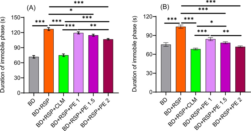
Assessment of behavioral test. (A) Forced swimming test (FST), (B) Tail suspension test (TST). BD = Basal diet, BD + RSP = Basal diet + Reserpine, BD + RSP + CLM= Basal diet + Reserpine + Clomipramine, BD + RSP + PE 1 = Basal diet + Reserpine + Phyllanthus emblica 1 mL/kg, BD + RSP + PE 1.5 = Basal diet + Reserpine + Phyllanthus emblica 1.5 mL/kg, BD + RSP + PE 2 = Basal diet + Reserpine + Phyllanthus emblica 2 mL/kg. Data are shown as Mean ± SEM. Level of significance *p < 0.05, **p < 0.01, ***p < 0.001.
In the TST, the positive control group treated with clomipramine (Group 3) had the shortest immobility phase at 68.4 seconds, indicating the drug's effectiveness in reducing stress (Fig. 1B). In contrast, the negative control group (Group 2) had the longest immobility phase at 104 seconds (p < 0.05), reflecting higher stress levels. Mice who received different doses of amla exhibited a dose-dependent response, with Group 6 showing the highest reduction in stress, followed by Groups 5 and 4. This suggests that higher doses of amla are associated with greater stress reduction, highlighting the medicinal benefits of amla in managing stress-related behaviors.
Stress-induced damage to the pancreas can hinder its ability to produce insulin, the hormone responsible for regulating blood sugar levels. This leads to hyperglycemia (elevated blood sugar) due to the body’s inability to manage glucose properly, underscoring the importance of stress management and pancreatic health for overall metabolic function.21
Fig. 2 presents the FBG levels and MDA concentration of different treatment groups of mice. Both parameters were significantly different across groups (p < 0.001). The basal diet group (Group 1) exhibited a mean FBG level of 5.58 mmol/L, representing normal glycemic regulation. In contrast, the negative control group (Group 2), exposed to oxidative stress, demonstrated a significantly elevated FBG level of 6.30 mmol/L (p < 0.05), suggesting stress-induced impairment of glucose homeostasis. The positive control group (Group 3), treated with clomipramine, showed a significantly reduced FBG level of 4.58 mmol/L (p < 0.05) compared to basal diet and negative control group. The significant normalization of FBG and MDA levels in the clomipramine-treated group further validates the suitability of the reserpine-induced oxidative stress model and confirms its responsiveness to standard pharmacological intervention.22 Amla-treated groups (Groups 4, 5, and 6) exhibited a clear dose-dependent decrease in FBG levels, with mean values of 4.52, 4.32, and 4.14 mmol/L, respectively.
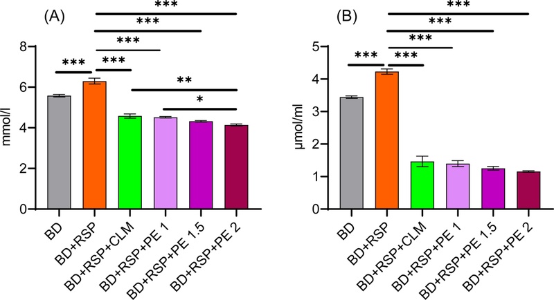
Variation of biochemical parameters among different groups. A. Fasting blood glucose, B. Malondialdehyde (MDA). BD = Basal diet, BD + RSP = Basal diet + Reserpine, BD + RSP + CLM = Basal diet + Reserpine + Clomipramine, BD + RSP + PE 1 = Basal diet + Reserpine + Phyllanthus emblica 1 mL/kg, BD + RSP + PE 1.5 = Basal diet + Reserpine + Phyllanthus emblica 1.5 mL/kg, BD + RSP + PE 2 = Basal diet + Reserpine + Phyllanthus emblica 2 mL/kg. Data are shown as Mean ± SEM. Level of significance *p < 0.05, **p < 0.01, ***p < 0.001.
To investigate the probable association between oxidative stress and body weight regulation, different groups of mice were subjected to distinct treatment conditions (Fig. 3). The negative control group (Group 2) exhibited the significantly lowest body weight gain (3.2 g, p < 0.05), indicating a potential impact of oxidative stress on metabolic function. In contrast, the basal diet group (Group 1) and the clomipramine-treated group (Group 3) showed higher gains of 7.0 g and 8.0 g, respectively. Among the amla-treated groups, a dose-dependent trend in weight gain was observed. Group 4 (1 mL/kg) gained 5.2 g, Group 5 (1.5 mL/kg) gained 6.2 g, and Group 6 (2 mL/kg) exhibited the highest gain at 7.6 g. These findings suggest that increasing doses of amla extract may mitigate the adverse effects of oxidative stress on weight regulation, supporting its potential role in stress alleviation and metabolic stability.
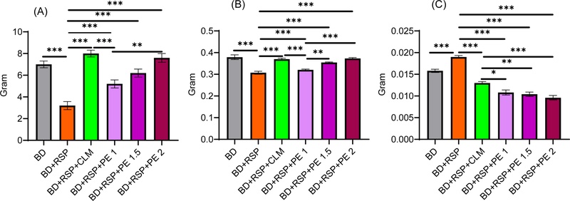
Variation of weight among different groups. (A) Body weight gain, (B) Brain weight, (C) Adrenal gland weight. BD = Basal diet, BD + RSP = Basal diet + Reserpine, BD + RSP + CLM = Basal diet + Reserpine + Clomipramine, BD + RSP + PE 1= Basal diet + Reserpine + Phyllanthus emblica 1 mL/kg, BD + RSP + PE 1.5 = Basal diet + Reserpine + Phyllanthus emblica 1.5 mL/kg, BD + RSP + PE 2 = Basal diet + Reserpine + Phyllanthus emblica 2 mL/kg. Data are shown as Mean ± SEM. Level of significance *p < 0.05, **p < 0.01, ***p < 0.001.
While the antioxidant activity of P. emblica in DPPH assays has been previously documented, the present study extends beyond confirmatory in vitro findings. The novelty of this work lies in integrating antioxidant profiling with a validated reserpine-induced oxidative stress model and assessing dose-dependent behavioral and biochemical responses. This combined approach enables a more functional interpretation of antioxidant capacity, linking phytochemical content to physiological stress modulation rather than relying solely on chemical radical scavenging assays. Amla is packed with high vitamin C content along with a variety of bioactive compounds, including polyphenols (flavonoids, tannins, gallic acid, ellagic acid), alkaloids, terpenoids, etc. which collectively contribute to its pronounced antioxidant properties.7 This elevated phenolic content underscores the presence of bioactive antioxidant constituents within the extract, which are widely recognized for their role in enhancing immune function and mitigating oxidative stress. The present findings are consistent with a previous study by Dasgupta documented the phytochemical composition of Indian-grown Phyllanthus emblica and reported slightly lower total phenolic content of 130.33 mg GAE/100 g, suggesting a general consistency in the antioxidant profile of amla across diverse geographical origins.23 However, a notable variation was observed when compared to the results of Karpagavalli who reported a markedly higher total polyphenol content of 2904.00 ± 9.22 mg GAE/100 g.24 This disparity is likely attributable to variations in sample preparation, extraction solvents, and particularly the sensitivity and calibration standards used in the spectrophotometric assays.25–27 Supporting the above-mentioned variability, Singh also reported elevated TPC values in amla seeds, suggesting that different plant parts and extraction targets can influence phenolic yield.27 In contrast, Madhavi observed a lower TPC value of approximately 112.5 mg GAE/100 g in their herbal formulations incorporating amla, which may be attributed to dilution effects or loss of active compounds during formulation.25 Furthermore, studies by Chaudhary and Singh observed a wide range of TPC values depending on the solvent polarity, drying method, and plant maturity.26,28 These authors emphasized that methanolic and aqueous extracts tend to yield different TPC values due to the solubility of phenolic compounds, and that temperature and drying duration also affect the integrity of bioactive molecules. Collectively, the above comparisons highlight the importance of methodological standardization in antioxidant studies. While the current findings corroborate the antioxidant potential of amla as a moderate source of phenolic compounds, the significant variations reported across studies suggest that extraction efficiency, assay specificity, and plant source (e.g., region, ripeness, and plant part) critically determine the phenolic yield. Future studies should aim to harmonize these variables to enable more meaningful cross-study comparisons.
Similar results have been observed in an earlier study of Firdous, which also reported comparable IC50 values.29 However, other studies conducted in India have observed lower IC50 values for P. emblica, such as 32 μg/mL by Madhavi, 25 μg/mL by Patil and Killedar, and 42 μg/mL by Singh, suggesting slightly higher antioxidant potency in those extracts.17,25,27 Conversely, some studies have reported higher IC50 values, such as 180.35 μg/mL and 290 μg/mL indicating weaker activity.26,28 These variations can be attributed to differences in extraction methods, solvent polarities, plant part used, and environmental or geographical factors affecting the phytochemical composition of the fruit.25–27
Overall, the highest immobility in the negative control group and the decrease of immobility in a dose-dependent manner in the amla-treated groups supported its role in stress mitigation and behavioral improvement in mice. Similar observations have been reported in studies conducted in India by Sohal and Dhingra, where amla and its derivatives significantly decreased immobility times in both FST and TST, highlighting its potential as a natural therapeutic for managing stress and depression-like behavior.30,31 The trend observed in the present study is consistent with the findings of Vasudevan and Parle, who also identified notable central nervous system benefits from amla fruit extracts.32 Importantly, no contradictory findings have been reported for amla in these behavioral assessments, reinforcing its role as a promising candidate for stress mitigation. The strong, dose-dependent reduction in immobility observed here, especially at the highest dose (Group 6), adds further evidence supporting the efficacy of amla as an adaptogenic and antidepressant agent, aligning with and extending the existing literature on its therapeutic benefits. Collectively, the significant reduction in immobility time in both FST and TST, particularly at higher doses of amla extract, reflects attenuation of reserpine-induced depressive-like behavior. Since reserpine induces oxidative stress-mediated monoamine depletion and neuronal dysfunction, the observed behavioral recovery suggests that P. emblica exerts neuroprotective effects, possibly through its antioxidant-mediated stabilization of neuronal integrity and neurotransmitter balance.
The reserpine treatment in mice resulted in an elevation of fasting blood glucose.33 Compared to the reserpine group, the amla-treated groups had significantly lower FBG levels, highlighting amla’s potential therapeutic role in reducing oxidative stress and improving glucose metabolism. The dose-response relationship reinforces the idea that higher doses of amla may offer greater protection against hyperglycemia caused by oxidative stress. These above findings are corroborated by earlier studies that underline the antidiabetic efficacy of amla. Rao demonstrated that amla extract significantly reduced hyperglycemia and oxidative markers in streptozotocin-induced diabetic rats, suggesting its role in bolstering antioxidant defenses and preserving pancreatic β-cell function.34 Similarly, another study reported the glucose lowering capacity of amla extract.35 In another investigation, Singh confirmed that amla administration (500 mg/kg) ameliorated arsenic-induced hyperglycemia by alleviating oxidative stress and restoring insulin secretion, further supporting its antidiabetic and cytoprotective properties.36
High serum levels of malondialdehyde (MDA) are a marker of oxidative stress, resulting from free radicals attacking the body’s lipids, leading to lipid peroxidation and MDA production.21 The mean serum MDA levels in this study provide insight into the potential antioxidant effects of the treatments. The clomipramine-treated positive control group (Group 3) showed a significant reduction in mean serum MDA levels (1.465 μmol/mL, p < 0.05) compared to both the negative control and basal diet groups, suggesting that clomipramine may possess antioxidant properties and help alleviate oxidative stress in mice. In the groups treated with different doses of amla extract (Groups 4, 5, and 6), a dose-dependent decrease in serum MDA levels was observed, with levels dropping consistently as the dose increased: 1.398 μmol/mL at 1 mL/kg, 1.254 μmol/mL at 1.5 mL/kg, and 1.157 μmol/mL at 2 mL/kg. This suggests that higher doses of amla have a stronger impact on reducing oxidative stress markers. Additionally, the effectiveness of amla in lowering MDA levels was found to be comparable to that of clomipramine, indicating its potential as a natural antioxidant and a means to reduce oxidative stress. These results align closely with a prior study by Golechha reported that amla extract significantly decreased elevated MDA levels caused by scopolamine-induced oxidative stress in the brain, indicating its neuroprotective role through enhanced antioxidant defense.37 Similarly, Singh observed that amla extract mitigated arsenic-induced lipid peroxidation, reducing MDA accumulation by approximately 20% relative to arsenic-only treatments, reinforcing its role as a robust natural antioxidant.38 Collectively, the above findings emphasize the significance of amla as a potent natural antioxidant and antidiabetic agent that can effectively mitigate oxidative stress-induced disturbances.
Oxidative stress is a state marked by an imbalance between the generation of reactive oxygen species (ROS) and the biological capacity to neutralize their effects, potentially leading to cellular and mitochondrial damage. This disruption is especially significant for energy metabolism, as mitochondria are highly susceptible to ROS-induced impairments that can reduce ATP generation and compromise cellular metabolism.39 Furthermore, it might affect the body’s ability to regulate hunger and metabolism because it can increase inflammation and disrupt metabolic functions.40 The current observations in the weight test are in agreement with prior studies highlighting amla’s efficacy in counteracting ROS-induced metabolic disturbances. For instance, Rao and Gouda demonstrated that amla extract mitigated oxidative damage and promoted weight recovery in rodent models.34,41 However, inconsistencies have been observed in certain toxicological studies. Fazal reported that amla administered in isolation was insufficient to restore body weight in rats exposed to arsenic and lead toxicity, although its efficacy was enhanced when combined with ginger.42 Similarly, Muthu observed that in a hypothyroidism and high-fat diet model, amla significantly improved oxidative stress markers but failed to produce a notable effect on body weight, suggesting that comorbid endocrinopathies and dietary factors can modulate the outcomes of amla therapy.43 Together, these findings underscore amla’s role as a potent antioxidant and metabolic modulator while highlighting the nuanced interplay between oxidative stress, physiological context, and amla’s therapeutic efficacy.
The brain, due to its high oxygen content and rapid metabolism, is particularly vulnerable to oxidative stress, especially in the hippocampus, which is vital for memory and learning.39 Oxidative stress can inhibit neuron production, damage existing neurons, disrupt communication between them, and increase inflammation.44 The impact of oxidative stress and the therapeutic efficacy of various interventions on brain weight were assessed, as presented also in Table 5. Mice in the basal diet group (Group 1) exhibited the highest mean brain weight (0.379 g), whereas the negative control group (Group 2), subjected to induced oxidative stress, showed a marked reduction to 0.307 g. Similar findings have been reported by Singh, who observed marked decreases in brain protein content and associated structural deterioration in gamma-irradiated mice, highlighting the vulnerability of brain tissue to oxidative damage.45
The positive control group (Group 3), treated with clomipramine, demonstrated a mean brain weight of 0.370 g, suggesting partial neuroprotection. Amla-treated groups displayed a dose-dependent increase in brain weight: Group 4 (0.320 g), Group 5 (0.354 g), and Group 6 (0.373 g). Notably, Group 6, which received the highest dose of P. emblica extract (2 mL/kg), exhibited a brain weight comparable to that of the basal diet group and surpassed that of the clomipramine group. These findings suggest that higher doses of amla may exert neuroprotective effects, potentially counteracting oxidative stress-induced reductions in brain mass. This finding is also consistent with Singh, who observed that amla extract mitigated gamma radiation-induced brain damage, preserving both structural and biochemical integrity.45 The neuroprotective potential of amla has been attributed to its rich polyphenolic composition and antioxidant activity, which neutralize free radicals and maintain cellular homeostasis.46
However, not all studies have confirmed such benefits. Gouda reported no significant changes in brain weight following amla treatment.41 This discrepancy may be due to differences in extract preparation and bioactive compound profiles.25–27 The study by Gouda used an ethyl acetate fraction, which may lack the higher concentrations of polar phenolic constituents present in the methanolic extract used in the current study, thereby affecting its efficacy.41 Overall, these findings underscore the potential of amla as a neuroprotective agent, highlighting its ability to counteract oxidative stress-induced structural changes in the brain. The restoration of brain weight in amla-treated groups, particularly at 2 mL/kg, represents a key finding of this study. Reserpine-induced oxidative stress significantly reduced brain mass, reflecting potential neuronal loss or structural compromise. The dose-dependent reversal of this reduction suggests that P. emblica extract preserves neural tissue integrity under oxidative stress conditions. When considered alongside reduced MDA levels and improved behavioral outcomes, these findings collectively provide convergent evidence of neuroprotection. Thus, the neuroprotective effect of amla, demonstrated through behavioral recovery, reduced lipid peroxidation, and preservation of brain morphology, constitutes a primary contribution of this work.
The regulation of cortisol, a key stress hormone, is controlled by a feedback loop between the hypothalamus, pituitary gland, and adrenal glands, known as the hypothalamic-pituitary-adrenal (HPA) axis. Oxidative stress can disrupt this loop, leading to abnormal cortisol secretion.47 One outcome of increased oxidative stress is adrenal hypertrophy, or enlargement of the adrenal glands, which correlates with higher cortisol production.15 The influence of oxidative stress on adrenal gland hypertrophy and the modulatory effects of various treatments were evaluated. The basal diet (Group 1) exhibited a mean adrenal gland weight 0.016 g, serving as the physiological reference. The negative control group (Group 2), subjected to oxidative stress via reserpine administration, showed a significant increase in adrenal weight to 0.019 g (p < 0.05), suggesting stress-induced adrenal hyperplasia.
In contrast, the clomipramine-treated positive control group (Group 3) demonstrated a significantly reduced mean adrenal weight of 0.013 g (p < 0.05), indicating its potential to mitigate stress responses. Notably, the amla-treated groups displayed a dose-dependent decrease in adrenal weight in Groups 4, 5, and 6. This inverse correlation between amla dosage and adrenal gland weight supports the hypothesis that P. emblica confers protective effects against oxidative stress, potentially normalizing adrenal morphology in a manner comparable to pharmacological interventions. These findings underscore the adaptogenic potential of P. emblica, aligning with earlier observations by Neelima, who reported significant decreases in adrenal gland weight and corticosterone levels following amla administration in a rodent stress model.25 Although some studies have examined the antioxidant and adaptogenic benefits of amla in animal models , none have contradicted its efficacy in mitigating stress-induced adrenal hypertrophy.48,49 Together, these findings highlight the role of amla as a promising natural modulator of HPA axis activity, reinforcing its potential in the prevention and management of oxidative stress-associated endocrinopathies.
Although the present study quantified total phenolic content and evaluated antioxidant activity, detailed phytochemical profiling using HPLC-UV or LC-MS was not performed. Therefore, the specific major and minor constituents responsible for the observed biological effects cannot be definitively identified. Future studies incorporating chromatographic and mass spectrometric analyses are warranted to standardize the extract and correlate individual bioactive compounds with pharmacological activity.
In conclusion, the present study demonstrates that Bangladeshi-grown Phyllanthus emblica possesses significant antioxidant and neuroprotective properties in a reserpine-induced oxidative stress model. The extract not only improved glycemic parameters but also restored brain weight and attenuated depressive-like behavior in a dose-dependent manner. These findings position amla as a promising natural neuroprotective agent for oxidative stress-associated neurological conditions, warranting further mechanistic and translational investigations.
Acknowledgments
We extend our thanks to our diligent lab assistant, Md Mahbub Alam, whose invaluable assistance, and cooperation have facilitated the smooth execution of experiments and various aspects of this research. Additionally, we are indebted to the Pharmacy Department for granting us access to their mice lab and allowing us to work with their facilities and utilities, which significantly enriched the scope of our study.
Conflicts of Interest
The authors of this article declare that they have no conflicts of interest.
References
-
Sharifi-Rad, M.; Kumar, N. V. A.; Zucca, P.; Varoni, E. M.; Dini, L.; Panzarini, E.; Rajkovic, J.; Fokou, P. V. T.; Azzini, E.; Peluso, I.; Mishra, A. P.; Nigam, M.; Rayess, Y. E.; Beyrouthy, M. E.; Polito, L.; Iriti, M.; Martins, N.; Martorell, M.; Docea, A. O.; Setzer, W. N.; Calina, D.; Cho, W. C.; Sharifi-Rad, J. Front. Physiol. 2020, 11, 694.
[https://doi.org/10.3389/fphys.2020.00694]

-
Rahaman, M. M.; Hossain, R.; Herrera-Bravo, J.; Islam, M. T.; Atolani, O.; Adeyemi, O. S.; Owolodun, O. A.; Kambizi, L.; Daştan, S. D.; Calina, D.; Sharifi-Rad, J. Food Sci. Nutr. 2023, 11, 1657–1670.
[https://doi.org/10.1002/fsn3.3217]
-
Delgado, A. M.; Issaoui, M.; Chammem, N. J. AOAC Int. 2019, 102, 1356–1364.
[https://doi.org/10.5740/jaoacint.19-0128]
-
Zhang, H.; Tsao, R. Curr. Opin. Food Sci. 2016, 8, 33–42.
[https://doi.org/10.1016/j.cofs.2016.02.002]
-
Hussain, T.; Tan, B.; Yin, Y.; Blachier, F.; Tossou, M. C. B.; Rahu, N. Oxid. Med. Cell. Longev. 2016, 2016, 7432797.
[https://doi.org/10.1155/2016/7432797]
-
Rahman, M. M.; Ferdous, K. U.; Roy, S.; Nitul, I. A.; Mamun, F.; Hossain, M. H.; Subhan, N.; Alam, M. A.; Haque, M. A. Food Sci. Nutr. 2020, 8, 3578–3589.
[https://doi.org/10.1002/fsn3.1640]
-
Golechha, M.; Sarangal, V.; Ojha, S.; Bhatia, J.; Arya, D. S. Int. J. Inflam. 2014, 2014, 178408.
[https://doi.org/10.1155/2014/178408]
-
Baliga, M. S.; Dsouza, J. J. Eur. J. Cancer Prev. 2011, 20, 225–239.
[https://doi.org/10.1097/CEJ.0b013e32834473f4]
-
Yokozawa, T.; Kim, H. Y.; Kim, H. J.; Okubo, T.; Chu, D.-C.; Juneja, L. R. Br. J. Nutr. 2007, 97, 1187–1195.
[https://doi.org/10.1017/S0007114507691971]
-
Singh, M. K.; Yadav, S. S.; Yadav, R.S.; Chauhan, A.; Katiyar, D.; Khattri, S. Springerplus 2015, 4, 438.
[https://doi.org/10.1186/s40064-015-1227-9]
-
Hossain, M. M.; Hamza, A.; Polash, S. A.; Tushar, M. H.; Takikawa, M.; Piash, A. B.; Dekiwadia, C.; Saha, T.; Takeoka, S.; Sarker, S. R. RSC Pharm. 2024, 1, 245–258.
[https://doi.org/10.1039/D3PM00077J]
-
Sapkota, B. K.; Khadayat, K.; Sharma, K.; Raut, B. K.; Aryal, D.; Thapa, B. B.; Parajuli, N. Adv. Pharmacol. Pharm. Sci. 2022, 4929824.
[https://doi.org/10.1155/2022/4929824]
-
Ainsworth, E. A.; Gillespie, K. M. Nat. Protoc. 2007, 2, 875–877.
[https://doi.org/10.1038/nprot.2007.102]
- Molyneux, P. Songklanakarin J. Sci. Technol. 2004, 26, 211–219.
-
Wilson, C. B.; McLaughlin, L. D.; Nair, A.; Ebenezer, P. J.; Dange, R.; Francis, J. PLoS One, 2013, 8, e76146.
[https://doi.org/10.1371/journal.pone.0076146]
-
Lee, J.-S.; Kang, J.-Y.; Son, C.-G. Front. Behav. Neurosci. 2021, 14, 616389.
[https://doi.org/10.3389/fnbeh.2020.616389]
-
Patil, P.; Killedar, S. Heliyon 2021, 7, e06526.
[https://doi.org/10.1016/j.heliyon.2021.e06526]
- Sun, C.; Li, X.; Liu, L.; Canet, M. J.; Guan, Y.; Fan, Y.; Zhou, Y. Int. J. Clin. Exp. Med. 2016, 9, 4186–4189.
-
Adegbola, M. V.; Anyim, G.; Ntwasa, M.; Ayeleso, A. O.; Oyedepo, T. A. Appl. Sci. 2022, 12, 8216.
[https://doi.org/10.3390/app12168216]
-
Sleigh, J. N.; Weir, G. A.; Schiavo, G. BMC Res. Notes 2016, 9, 82.
[https://doi.org/10.1186/s13104-016-1915-8]
-
Ito, F.; Sono, Y.; Ito, T. Antioxidants 2019, 8, 72.
[https://doi.org/10.3390/antiox8030072]
-
Miftah, Z. F.; Khanam, J.; Mahadi, S. F. A.; Islam, S.N. Food Sci. Nutr. Res. 2021, 4, 1–7.
[https://doi.org/10.33425/2641-4295.1041]
-
Dasgupta, S.; Pandya, M.; Patel, N. Int. J. Curr. Pharm. Res. 2017, 9, 28–31.
[https://doi.org/10.22159/ijcpr.2017v9i5.22132]
-
Karpagavalli, B.; Amutha S.; Padmini, T.; Palanisamy R.; Chandrakumar, K. Indian J. Sci. Technol. 2014, 4, 672–677.
[https://doi.org/10.17485/ijst/2014/v7i5.3]
-
Madhavi, N.; Kumar, D.; Naman, S.; Singh, M.; Singh, P. A.; Bajwa, N.; Baldi, A. J. Drug Deliv. Ther. 2019, 9, 212–221.
[https://doi.org/10.22270/jddt.v9i4.3029]
-
Chaudhary, N.; Sabikhi, L.; Hussain, S. A.; M. H. Kumar, S. J. Herb. Med. 2020, 22, 100343.
[https://doi.org/10.1016/j.hermed.2020.100343]
- Singh, P. G.; Jain, A. S.; Setty, P. B. S.; Bv, S.; Patil, S. S.; Ts, G.; Suresh, K. P.; Dugganaboyana, G. K.; Murugesan, K.; Gnanasekaran, A.; Shivamallu, C.; Kollur, S. P.; Srinivasa, C.; HI, R.; Rudrapathy, P.; Basalingappa, K. M. Bioinformation 2022, 18, 683–691.
-
Singh, R.; Kumar, S. J. Drug Deliv. Ther. 2021, 11, 32–35
[https://doi.org/10.22270/jddt.v11i1.4667]
-
Firdous, A.; Ringø, E.; Elumalai, P. Aquac. Fish 2020, 6, 617–627.
[https://doi.org/10.1016/j.aaf.2020.09.003]
- Sohal, A.; Joshi, S. S.; Koli, P. G.; Puradkar, P. V.; Date, V. C. Cureus 2024, 16, e72859.
-
Dhingra, D.; Joshi, P.; Gupta, A.; Chhillar, R. CNS Neurosci. Ther. 2011, 18, 419–425.
[https://doi.org/10.1111/j.1755-5949.2011.00256.x]
-
Vasudevan, M.; Parle, M. Physiol. Behav. 2007, 91, 46–54.
[https://doi.org/10.1016/j.physbeh.2007.01.016]
-
Soliman, E.; Essmat, N.; Mahmoud, M. F.; Mahmoud, A. A. A. Naunyn Schmiedebergs Arch. Pharmacol. 2020, 393, 1391–1404.
[https://doi.org/10.1007/s00210-020-01838-w]
-
Rao, T. P.; Sakaguchi, N.; Juneja, L. R.; Wada, E.; Yokozawa, T. J. Med. Food 2005, 8, 362–368.
[https://doi.org/10.1089/jmf.2005.8.362]
- Huang, S.-M.; Lin, C.-H.; Chang, W.-F.; Shih, C.-C. Food Nutr. Res. 2023, 67, 9854.
-
Singh, M. K.; Dwivedi, S.; Yadav, S. S.; Yadav, R. S.; Khattri, S. Indian J. Clin. Biochem. 2020, 35, 179–187.
[https://doi.org/10.1007/s12291-019-00820-5]
- Golechha, M.; Bhatia, J.; Arya, D. S. J. Environ. Biol. 2012, 33, 95–100.
-
Singh, M. K.; Dwivedi, S.; Yadav, S. S.; Sharma, P.; Khattri, S. Indian J. Clin. Biochem. 2014, 29, 29–37.
[https://doi.org/10.1007/s12291-013-0353-9]
-
Cenini, G.; Lloret, A.; Cascella, R. Oxid. Med. Cell. Longev. 2019, 2019, 2105607.
[https://doi.org/10.1155/2019/2105607]
-
Tang, H.; Inoki, K.; Brooks, S. V.; Okazawa, H.; Lee, M.; Wang, J.; Kim, M.; Kennedy, C. L.; Macpherson, P. C. D.; Ji, X.; Van Roekel, S.; Fraga, D. A.; Wang, K.; Zhu, J.; Wang, Y.; Sharp, Z. D.; Miller, R. A.; Rando, T. A.; Goldman, D.; Guan, K.-L.; Shrager, J. B. Aging Cell 2019, 18, e12943.
[https://doi.org/10.1111/acel.12943]
-
Gouda, B.; Sinha, S. N.; Sangaraju, R.; Huynh, T.; Patangay, S.; Mullapudi, S. V.; Mungamuri, S. K.; Patil, P. B.; Periketi, M. C. Heliyon 2024, 10, e34992.
[https://doi.org/10.1016/j.heliyon.2024.e34992]
-
Fazal, M.; Veeraraghavan, V. P.; Tahreen, B.; Jayaraman, S.; Gayathri, R. Bioinformation 2021, 17, 295–305.
[https://doi.org/10.6026/97320630017295]
-
Muthu, P. R.; Bobby, Z.; Sankar, P.; Vickneshwaran, V.; Jacob, S. E. J. Basic. Clin. Physiol. Pharmacol. 2017, 29, 175–184.
[https://doi.org/10.1515/jbcpp-2017-0116]
-
Hajipour, S.; Farbood, Y.; Gharib-Naseri, M. K.; Goudarzi, G.; Rashno, M.; Maleki, H.; Bakhtiari, N.; Nesari, A.; Khoshnam, S. E.; Dianat, M.; Sarkaki, B.; Sarkaki, A. Life Sci. 2019, 242, 117210.
[https://doi.org/10.1016/j.lfs.2019.117210]
-
Singh, I.; Sharma, A.; Jindal, A.; Soyal, D.; Goyal, P. K. Ann. Neurosci. 2010, 13, 65–71.
[https://doi.org/10.5214/ans.0972.7531.2006.130302]
-
Gul, M.; Liu, Z.-W.; Iahtisham-Ul-Haq; Rabail, R.; Faheem, F.; Walayat, N.; Nawaz, A.; Shabbir, M. A.; Munekata, P. E. S.; Lorenzo, J. M.; Aadil, R. M. Antioxidants 2022, 11, 816.
[https://doi.org/10.3390/antiox11050816]
-
Shal, B.; Khan, A.; Naveed, M.; Khan, N. U.; Ihsan-Ul-Haq; AlSharari, S. D.; Kim, Y. S.; Khan, S. Biomed. Pharmacother. 2019, 111, 209–223.
[https://doi.org/10.1016/j.biopha.2018.12.047]
-
Rege, N. N.; Thatte, U. M.; Dahanukar, S. A. Phytother. Res. 1999, 13, 275–291.
[https://doi.org/10.1002/(SICI)1099-1573(199906)13:4<275::AID-PTR510>3.0.CO;2-S]
-
Panda, S.; Kar, A. Pharmazie 2003, 58, 753–755.
[https://doi.org/10.1037/0003-066X.58.9.753]