
Anti-inflammatory Activity of Compounds from the Aerial Part of Perilla frutescens var. acuta (Thunb.) Kudo
Abstract
A new depside glucoside, perilloside F (1), together with fifteen known compounds (2–16), was isolated from the aerial parts of Perilla frutescens var. acuta (Thunb.) Kudo (Labiatae). The chemical structures of all compounds were elucidated using spectroscopic methods, including 1D and 2D NMR spectroscopy and high-resolution electrospray ionization spectrometry (HR-ESI-MS), as well as by comparison with previously published data. All isolated compounds were evaluated for their anti-inflammatory activity by assessing their inhibitory effects on nitric oxide (NO) production in RAW 264.7 macrophage cells. Six compounds (3, 5, 8, 9, 11, and 13) exhibited significant inhibition of NO production, with IC50 values of 65.72, 15.35, 17.31, 10.55, 12.31, and 39.01 μM, respectively. Among them, compound 9 demonstrated the strongest inhibitory activity, with an IC50 value of 10.55 μM. Importantly, none of the active compounds exhibited cytotoxicity at concentrations below 50 μM.
Keywords:
Perilla frutescens, Labiatae, Anti-inflammatory, NO productionIntroduction
Perilla frutescens var. acuta (Thunb.) Kudo is an annual plant belonging to the Labiatae family. It is widely distributed throughout Asia, particularly in China, Japan, Korea, and Vietnam. Historically, P. frutescens has played a significant role in the traditional medicinal systems of various Asian countries. It has long been used as a natural remedy for a wide range of conditions, including depression, asthma, anxiety, tumors, coughs, allergies, intoxication, common colds, fever, chills, headaches, nasal congestion, and gastrointestinal disorders. The rich phytochemical composition of P. frutescens contributes to its diverse applications. The plant contains a variety of bioactive compounds, including terpenoids and phenolic compounds such as flavonoids. Previous biological studies have demonstrated that P. frutescens exhibited a broad spectrum of biological activities, including anti-inflammatory, anti-depressant, anticancer, antioxidant, antimicrobial, insecticidal, neuroprotective, and hepatoprotective effects.1,2 Notably, Perilla frutescens leaf extract was reported to suppress the mRNA expression and protein production of pro-inflammatory mediators, such as extracellular signal regulated kinase (ERK)1/2, c-Jun N-terminal kinase (JNK), p38, and NF-κB signaling in RAW 264.7 cells stimulated with LPS.3
Nitric oxide (NO) plays an important physiological role by exerting cytoprotective effects, regulating vascular relaxation, inhibiting platelet aggregation and leukocyte adhesion, and modulating cytokine production. However, excessive or chronic NO production (primarily mediated by the inducible NOS) can shift its role to that of a pro-inflammatory mediator, resulting in toxic effects. This excess NO can react with reactive oxygen species (ROS) to form reactive nitrogen species (RNS). The overproduction of RNS may lead to nitrosative stress, which can damage proteins, lipids, and DNA, potentially leading to apoptosis (programmed cell death). Because NO production plays a critical role in the inflammatory process, modulation of NO levels (through inhibition of iNOS activity or suppression of iNOS protein expression) represents a promising strategy for assessing the anti-inflammatory potential of various compounds.3–5 In the present study, we carried out the isolation and structural determination of compounds from the aerial parts of P. frutescens, as well as evaluated their anti-inflammatory effects by assessing LPS-induced NO production in RAW 264.7 macrophage cells.
Materials and Methods
General experimental procedures − Reverse-phase RP-C18 (75 μm, Merck, Germany) and normal-phase silica gel (40–63 μm and 63–200 μm, Merck, Germany) were used for open column chromatography. Merck precoated silica gel RP-C18 F254 and 60 F254 plates were used to perform TLC. A Thermo spectrophotometer recorded UV spectra, while the IR spectrum was measured using a JASCO FFT/IR-4100 spectrometer. The NMR spectra were obtained using a Bruker Avance 500 MHz NMR spectrometer (Bruker, MA, USA) and a Varian Unity Inova 400 MHz (Varian, Inc., California, USA). High-resolution electrospray ionization-mass spectrometry (HR-ESI-MS) was measured on a SYNAPT G2 (Waters, U.K.) mass spectrometer. A TECAN Infinite F200 Pro Microplate Reader was used to calculate the bioactivity test.
Plant material − Perilla frutescens var. acuta aerial parts were collected in November 2023 from the medicinal plant garden at the College of Pharmacy, Daegu Catholic University, Republic of Korea (35°54′43.3″ N, 128°48′16.4″ E). The specimen was identified by Professor Byung Sun Min, and the voucher specimen, CUD-2634-1, has been deposited at the College of Pharmacy, Daegu Catholic University, Republic of Korea.
Chemicals and Reagents − All solvents were purchased from Daejung Chemicals and Metals Company (Korea) and Duksan Pure Chemicals Company (Korea). The macrophage cell line RAW 264.7 was obtained from the Korean Cell Line Bank (Korea). DPBS was purchased from GIBCO (USA), and DMEM and penicillin–streptomycin were bought from CYTIVA (USA). FBS was bought from ATLAS Biologicals (USA), and LPS was purchased from Sigma-Aldrich (USA).
Extraction and isolation − The aerial parts of P. frutescens (7.5 kg) were extracted with MeOH (15 L × 3 times) under reflux for 3 hours. This process yielded crude extract, which was then filtered and evaporated in the solvent under reduced pressure, resulting in 853 g of the MeOH extract. The MeOH extract was further suspended in hot water (2 L) and partitioned with different solvents, including n-hexane (2 L × 3 times), CH2Cl2 (2 L × 3 times), and EtOAc (2 L × 3 times), to obtain n-hexane (114.5 g), CH2Cl2 (91.03 g), EtOAc (97.77 g) fractions, and water fraction, respectively.
The CH2Cl2 fraction (91.03 g) underwent silica gel column chromatography, and a gradient of CH2Cl2:acetone (19/1–1/4, v/v) was used for elution, resulting in ten fractions (MC1–MC10). Fraction MC6 (1.7 g) underwent repeated silica gel column chromatography, using CH2Cl2:acetone (50/1–1/1, v/v) as a solvent system to yield compound 12 (10 mg). Fraction MC7 (12.7 g) was fractionated by silica gel column chromatography with a gradient of CH2Cl2:acetone (9/1–1/4, v/v), and then purified by RP-C18 column chromatography (MeOH:water, 1:2–9:1) and crystallization to obtain compound 5 (7 mg), 8 (15 mg), 13 (12 mg), 14 (84 mg), and 15 (498 mg). A similar procedure was applied to fraction MC9 (6.5 g) to obtain compounds 6 (3 mg) and 7 (4 mg).
The EtOAc fraction (97.77 g) was chromatographed on silica gel, with a gradient of CH2Cl2:acetone (19/1–1/4, v/v), yielding eleven fractions (E1–E11). Fraction E11 (6.6 g) was fractionated by silica gel column chromatography with a gradient of CH2Cl2:acetone (4/1–1/4, v/v), producing eight subfractions (E11.1–E11.8). Subfractions E11.5 (710 mg), E11.6 (870 mg), and E11.7 (900 mg) were further purified by repeated RP-C18 silica gel column chromatography using methanol:water (1/9–1/4, v/v) to yield compound 1 (15 mg) and compound 2 (10 mg), respectively. Fraction E4 (4.0 g) was further fractionated by silica gel column chromatography using a mixture of CH2Cl2:acetone (9/1–1/4, v/v) as a solvent system, resulting in three subfractions (E4.1-E4.1). Subfraction E4.1 (750 mg) was then further purified on an RP-C18 silica gel column using a gradient of methanol: water (9/1–4/1, v/v), yielding compound 16 (68 mg). Fraction E7 (6.5 g) was fractionated by silica gel column chromatography with a gradient of CH2Cl2:acetone (9/1–1/4, v/v), producing seven subfractions (E7.1–E7.7). Subfraction E7.2 (150 mg) was chromatographed on a silica gel column using CH2Cl2:acetone (9/1, v/v) to yield compound 11 (30 mg). Subfraction E7.3 (1.6 g) was further fractionated by silica gel column chromatography, eluted with CH2Cl2:acetone (CH2Cl2:acetone, 9/1–1/1, v/v) to yield five subfractions (E7.3.1–E7.3.5). The subfraction E7.3.2 (300 mg) was purified further on an RP-C18 silica gel column chromatography, using methanol:water (1/3–1/1, v/v) as a solvent system to obtain compound 3 (27 mg) and compound 9 (28 mg). Subfraction E7.6 (150 mg) was further purified on an RP-C18 silica gel column using methanol:water (1/9–1/1, v/v), leading to the isolation of compound 4 (12 mg).
Water residue was run through the dianion column using a methanol:water solvent system (from 100% water, 25% methanol, 50% methanol, 75% methanol, and 100% methanol) to obtain 5 subfractions. Fraction W3 (33 g) was further fractionated by silica gel column chromatography using a solvent system of CH2Cl2:methanol (9/1–1/1, v/v), resulting in eleven subfractions (W3.1–W3.11). Subfraction W3.8 (1.2 g) was then further purified on an RP-C18 silica gel column using gradient methanol:water (9/1–1/1, v/v), yielding compound 10 (104 mg).
Perilloside F (1) – reddish amorphous solid; UV (MeOH) λmax nm: 269, 299; IR νmax (cm-1): 3332, 2941, 2830, 1454, 1417, 1112, 1024; 1H-NMR (500 MHz, pyridine-d5): δH 8.26 (1H, d, J = 2.0 Hz, H-2'), 7.99 (1H, dd, J = 8.3, 2.0 Hz, H-6'), 7.31 (1H, d, J = 8.3 Hz, H-5'), 7.29 (1H, d, J = 2.2 Hz, H-3), 7.05 (1H, d, J = 2.2 Hz, H-5), 5.49 (1H, d, J = 7.1 Hz, H-1"), 4.28–4.34 (5H, m, H-2", H-3", H-4", H-6"), 4.17(1H,d, J = 16.5 Hz, H-7a), 4.01 (1H, d, J = 16.5 Hz, H-7b), 3.89 (1H, m, H-5"), 3.49 (3H, s, H-9); 13C-NMR (125 MHz, pyridine-d5): δC 172.6 (C-8), 165.4 (C-7'), 159.7 (C-4), 158.7 (C-2), 153.7 (C-4'), 152.3 (C-6), 147.6 (C-3'), 124.2 (C-6'), 121.2 (C-1'), 118.5 (C-2'), 116.7 (C-5'), 109.2 (C-1), 105.6 (C-5), 103.6 (C-1"), 102.4 (C-3), 79.1 (C-5"), 78.8 (C-3"), 75.2 (C-2"), 71.2 (C-4"), 62.4 (C-6"), 52.0 (C-9), 30.2 (C-7); HR-ESI-MS m/z 519.1116 [M + Na]+ (calcd. for C22H24O13Na, 519.1115).
Hydrolysis of compound 1 – Compound 1 (2 mg) was dissolved in 3.0 mL of 10% HCl and then refluxed at 80°C for 3 hours. After cooling, the resulting mixture was extracted three times with 3 mL of ethyl acetate. The aqueous layers were evaporated to dryness with MeOH until neutral. Continuously, the residue was analyzed by TLC on silica gel (CHCl3:MeOH:H2O, 8:5:1) and compared with the standard sample (glucose, Rf = 0.32). According to the previously reported method, the sugar moiety was determined to be D-glucose.6
Indole-3-carboxylic acid (6) – White powder; 1H-NMR (500 MHz, methanol-d4): δ 8.19 (1H, dd, J = 7.6, 2.0 Hz, H-4), 7.79 (1H, s, H-2), 7.36 (1H, dd, J = 7.6, 1.7 Hz, H-7), 7.10 (2H, m, H-6 and H-5); 13C-NMR (125 MHz, methanol-d4): δ 174.6 (C-10), 138.0 (C-8), 131.1 (C-2), 128.2 (C-9), 122.6 (C-6), 122.5 (C-4), 121.2 (C-5), 114.7 (C-3), 112.3 (C-7).
Caryolane-1,9β-diol(7) – White amorphous powder; 1H-NMR (500 MHz, methanol-d4): δ 3.38 (1H, t, J = 3.4 Hz, H-9), 2.22 (1H, m, H-2), 2.02 (1H, m, H-10a), 1.89 (1H, ddd, J = 12.3, 8.5, 6.7 Hz, H-5), 1.73 (1H, ddt, J = 14.8, 5.6, 3.6 Hz, H-10b), 1.61 (1H, td, J = 12.2, 6.6 Hz, H-11a), 1.51 (2H, m, H-3), 1.48 (1H, m, H-6a), 1.45 (1H, m, H-11b), 1.40 (3H, m, H-12, H-7a), 1.36 (1H, m, H-6b), 1.11 (1H, m, H-7b), 0.99 (3H, s, H-14), 0.99 (3H, s, H-13), 0.88 (3H. s, H-15); 13C-NMR (125 MHz, methanol-d4): δ 72.6 (C-9), 71.5 (C-1), 44.9 (C-5), 43.3 (C-12), 40.0 (C-8), 39.6 (C-2), 36.6 (C-7), 35.6 (C-4), 35.1 (C-3), 33.9 (C-11), 30.8 (C-14), 28.8 (C-10), 27.3 (C-15), 21.4 (C-6), 21.3 (C-13).
trans-3,4,5-Trimethoxycinnamyl alcohol (8) – White powder; 1H-NMR (500 MHz, methanol-d4): δ 6.71 (2H, s, H-2, H-6), 6.53 (1H, d, J = 15.8 Hz, H-7), 6.31 (1H, dt, J = 15.8, 5.7 Hz, H-8), 4.22 (2H, dd, J = 5.7, 1.5 Hz, H-9), 3.84 (6H, s, 3-OCH3 and 5-OCH3), 3.75 (3H, s, 4-OCH3); 13C-NMR (125 MHz, methanol-d4): δ 154.6 (C-3 and C-5), 138.7 (C-4), 134.6 (C-1), 131.5 (C-7), 129.6 (C-8), 104.8 (C-2 and C-6), 63.6 (C-9), 61.1 (4-OCH3), 56.6 (3-OCH3 and 5-OCH3).
Luteolin (9) – Yellow amorphous powder; 1H-NMR (500 MHz, acetone-d6): δ 7.50 (1H, d, J = 2.0 Hz, H-2'), 7.47 (1H, dd, J = 8.3, 2.1 Hz, H-6'), 7.00 (1H, d, J = 8.3 Hz, H-5'), 6.58 (1H, s, H-3), 6.52 (1H, d, J = 2.0 Hz, H-6), 6.25 (1H, d, J = 2.0 Hz, H-8); 13C-NMR (125 MHz, acetone-d6): δ 183.1 (C-4), 165.1 (C-7), 164.9 (C-2), 163.4 (C-9), 158.8 (C-5), 150.1 (C-4'), 146.5 (C-3'), 123.8 (C-6'), 120.1 (C-1'), 116.6 (C-5'), 114.1 (C-2'), 105.3 (C-10), 104.2 (C-3), 99.7 (C-6), 94.7 (C-8).
Luteolin-5-O-glucoside (10) – Yellow amorphous powder; 1H-NMR (500 MHz, methanol-d4): δ 7.35 (1H, dd, J = 6.5, 2.2 Hz, H-6'), 7.34 (1H, d, J = 2.5 Hz, H-2'), 6.89 (1H, d, J = 8.9 Hz, H-5'), 6.81 (1H, d, J = 2.3, H-8), 6.69 (1H, d, J = 2.3, H-6), 6.52 (1H, s, H-3), 4.84 (1H, d, J = 7.7 Hz, H-1''), 3.95 (1H, dd, J = 12.0, 1.8 Hz, H-6a''), 3.76 (1H, dd, J = 12.0, 5.0 Hz, H-6b''), 3.60-3.48 (4H, m, H-2'', H-3'', H-4'', H-5'', sugar protons); 13C-NMR (125 MHz, methanol-d4): δ 180.4 (C-4), 164.8 (C-7), 164.4 (C-2), 160.6 (C-9), 160.1 (C-5), 150.8 (C-4'), 147.0 (C-3'), 123.5 (C-1'), 120.1 (C-6'), 116.8 (C-5'), 114.0 (C-2'), 109.3 (C-10), 106.5 (C-3), 105.0 (C-1''), 104.7 (C-6), 99.2 (C-8), 78.6 (C-5''), 77.3 (C-3''), 74.7 (C-2''), 71.2 (C-4''), 62.5 (C-6'').
Apigenin (11) – Yellow amorphous powder; 1H-NMR (500 MHz, methanol-d4): δ 7.85 (2H, d, J = 8.9 Hz, H-2' and H-6'), 6.93 (2H, d, J = 8.9 Hz, H-3' and H-5'), 6.59 (1H, s, H-3), 6.46 (1H, d, J = 2.1 Hz, H-8), 6.21 (1H, d, J = 2.1 Hz, H-6); 13C-NMR (125 MHz, methanol-d4): δ 183.9 (C-4), 166.3 (C-2), 166.1 (C-7), 163.2 (C-4'), 162.8 (C-5), 159.4 (C-9), 129.5 (C-2' and C-6'), 123.3 (C-1'), 117.0 (C-3' and C-5'), 105.3 (C-10), 103.8 (C-3), 100.1 (C-6), 95.1 (C-8).
3β-hydroxy-urs-11-en-28,13β-olide (12) – White powder; 1H-NMR (500 MHz, chloroform-d): δ 5.95 (1H, dd, J = 10.3, 1.0 Hz, H-12), 5.53 (1H, dd, J = 10.3, 3.1 Hz, H-11), 3.21 (1H, dd, J = 11.4, 4.8 Hz, H-3), 1.16 (3H, s, H-27), 1.05 (3H, s, H-26), 0.99 (3H, d, J = 6.4 Hz, H-29), 0.98 (3H, s, H-23), 0.93 (3H, d, J = 6.1 Hz, H-30), 0.91 (3H, s, H-25), 0.78 (3H, s, H-24); 13C-NMR (125 MHz, chloroform-d): δ 180.1 (C-28), 133.6 (C-12), 128.9 (C-11), 89.8 (C-13), 79.0 (C-3), 60.7 (C-18), 54.8 (C-5), 53.1 (C-9), 45.2 (C-17), 42.0 (C-8), 41.8 (C-14), 40.4 (C-20), 39.0 (C-4), 38.4 (C-1), 38.2 (C-19), 36.5 (C-10), 31.4 (C-22), 31.3 (C-7), 30.9 (C-21), 27.9 (C-24), 27.1 (C-2), 25.6 (C-15), 22.9 (C-16), 19.3 (C-30), 19.0 (C-26), 18.0 (C-25), 18.0 (C-29), 17.8 (C-6), 16.2 (C-27), 15.1 (C-23).
Betulinic acid (13) – White powder; 1H-NMR (500 MHz, methanol-d4): δ 4.69 (1H, d, J = 2.0 Hz, H-29), 4.57 (1H, dd, J = 2.1, 1.3 Hz, H-29), 3.12 (1H, dd, J = 11.2, 5.1 Hz, H-3), 3.00 (1H, td, J = 10.7, 4.6 Hz, H-19), 1.67 (3H, s, H-30), 0.98 (3H, s, H-27), 0.94 (3H, s, H-23), 0.93 (3H, s, H-26), 0.83 (3H, s, H-25), 0.74 (3H, s, H-24); 13C-NMR (125 MHz, methanol-d4): δ 179.9 (C-28), 151.5 (C-20), 110.1 (C-29), 79.4 (C-3), 57.1 (C-17), 56.4 (C-5), 51.6 (C-9), 50.1 (C-18), 48.0 (C-19), 43.3 (C-14), 41.6 (C-8), 39.8 (C-1), 39.6 (C-4), 39.3 (C-13), 38.0 (C-10), 37.9 (C-22), 35.2 (C-7), 33.1 (C-16), 31.4 (C-21), 30.5 (C-15), 28.5 (C-23), 27.7 (C-2), 26.5 (C-12), 21.8 (C-11), 19.6 (C-30), 19.1 (C-6), 16.6 (C-26), 16.5 (C-25), 16.0 (C-24), 15.1 (C-27).
Hyptadienic acid (14) – Colorless needles; 1H-NMR (500 MHz, methanol-d4): δ 5.38 (1H, brs, H-3), 5.28 (1H, brs, H-12), 4.19 (1H, dd, J = 14.8, 1.6 Hz, H-1a), 4.08 (1H, dd, J = 14.8, 1.4 Hz, H-1b), 1.30 (3H, s, H-27), 1.19 (3H, s, H-29), 1.12 (3H, s, H-25), 1.01 (3H, s, H-26), 0.93 (3H, s, H-23), 0.92 (3H, d, J = 6.8 Hz, H-30), 0.81 (3H, s, H-24); 13C-NMR (125 MHz, methanol-d4): δ 181.9 (C-28), 155.5 (C-2), 139.8 (C-13), 134.5 (C-3), 129.2 (C-12), 73.4 (C-19), 63.8 (C-5), 61.2 (C-1), 54.7 (C-18), 51.4 (C-10), 48.5 (C-17), 43.8 (C-20), 43.0 (C-9), 42.4 (C-8, C-14), 42.3 (C-4), 38.5 (C-22), 34.8 (C-7), 30.2 (C-23), 29.5 (C-15), 27.3 (C-29), 27.1 (C-16), 26.8 (C-11), 26.2 (C-21), 25.4 (C-27), 21.8 (C-24), 19.0 (C-25), 18.8 (C-26), 18.0 (C-6), 16.4 (C-30).
Inhibition of NO production and cell viability assay – The measurement of NO production was conducted by quantifying nitrite accumulation in the culture supernatant using the Griess reaction, following previously established methods. RAW 264.7 cells were treated with or without LPS (1 μg/mL) for 24 hours. After 1 day of incubation, 100 μL of Griess reagent was added to 100 μL of the cell medium, followed by the UV absorption at the wavelength of 570 nm. Cell viability assay was assessed using an MTT-based colorimetric assay. Each experiment was performed in triplicate, and IC50 values were calculated based on the average of three independent measurements.3
Results and Discussions
The methanol extract of the aerial part of P. frutescens var. acuta was sequentially partitioned into n-hexane, CH2Cl2, EtOAc, and aqueous fractions. Repeated column chromatography (silica gel, RP-C18) of the CH2Cl2, EtOAc, and aqueous fractions resulted in the isolation of one new depside compound (1) and fifteen known compounds (2–16). Based on the comprehensive spectroscopic analysis and comparison with published data, the fifteen known compounds were identified, including rosmarinic acid (2),7 methyl rosmarinate (3),8 caffeic acid (4),9 methyl caffeate (5),10 indole-3-carboxylic acid (6),11,12 caryolane-1,9β-diol (7),13 trans-3,4,5-trimethoxy cinnamyl alcohol (8),14 luteolin (9),15,16 luteolin-5-O-glucoside (10),8 apigenin (11),17,18 3β-hydroxy-urs-11-en-28,13β-olide (12),19 betulinic acid (13),20,21 hyptadienic acid (14),22,23 oleanolic acid (15),21 and ursolic acid (16).21
Compound 1 was isolated as a reddish amorphous solid. Its molecular formula was determined to be C22H24O13 by HR-ESI-MS, which showed a molecular ion peak at m/z 519.1116 [M + Na]+ (calculated for C22H24O13Na, 519.1115) (Fig. S1). The UV spectrum exhibited a peak at 269 nm with a shoulder at 299 nm, typical of a phenolic acid ester (Fig. S2). The 1H-NMR spectrum (Fig. S4) of 1 showed signals corresponding to several characteristic spin systems. One was characterized by an aromatic ABX system at δH 8.26 (1H, d, J = 2.0 Hz, H-2'), 7.99 (1H, dd, J = 8.3, 2.0 Hz, H-6'), and 7.31 (1H, d, J = 8.3 Hz, H-5'). Two meta-coupling aromatic protons at δH 7.29 (1H, d, J = 2.2 Hz, H-3) and 7.05 (1H, d, J = 2.2 Hz, H-5) indicated the presence of a 1,2,4,6-tetrasubstituted phenyl ring. In addition, a methylene group was observed at δH 4.17(1H, d, J = 16.5 Hz, H-7a) and 4.01 (1H, d, J = 16.5 Hz, H-7b), along with a methoxy group at δH 3.49 (3H, s, H-9). A sugar moiety was identified by the presence of an anomeric proton at δH 5.49 (1H, d, J = 7.1 Hz, H-1"), multiple peaks at δH 4.28–4.34 (5H, m, H-2", H-3", H-4", H-6", overlapping sugar protons), and a signal at δH 3.89 (1H, m, H-5"). Acid hydrolysis and subsequent TLC analysis of the corresponding monosaccharide derivative established the sugar moiety of 1 as ᴅ-glucose. The glucopyranosyl substituent was determined as β-configuration based on the large 3J1,2 coupling constant of the anomeric protons (J = 7.1 Hz).24
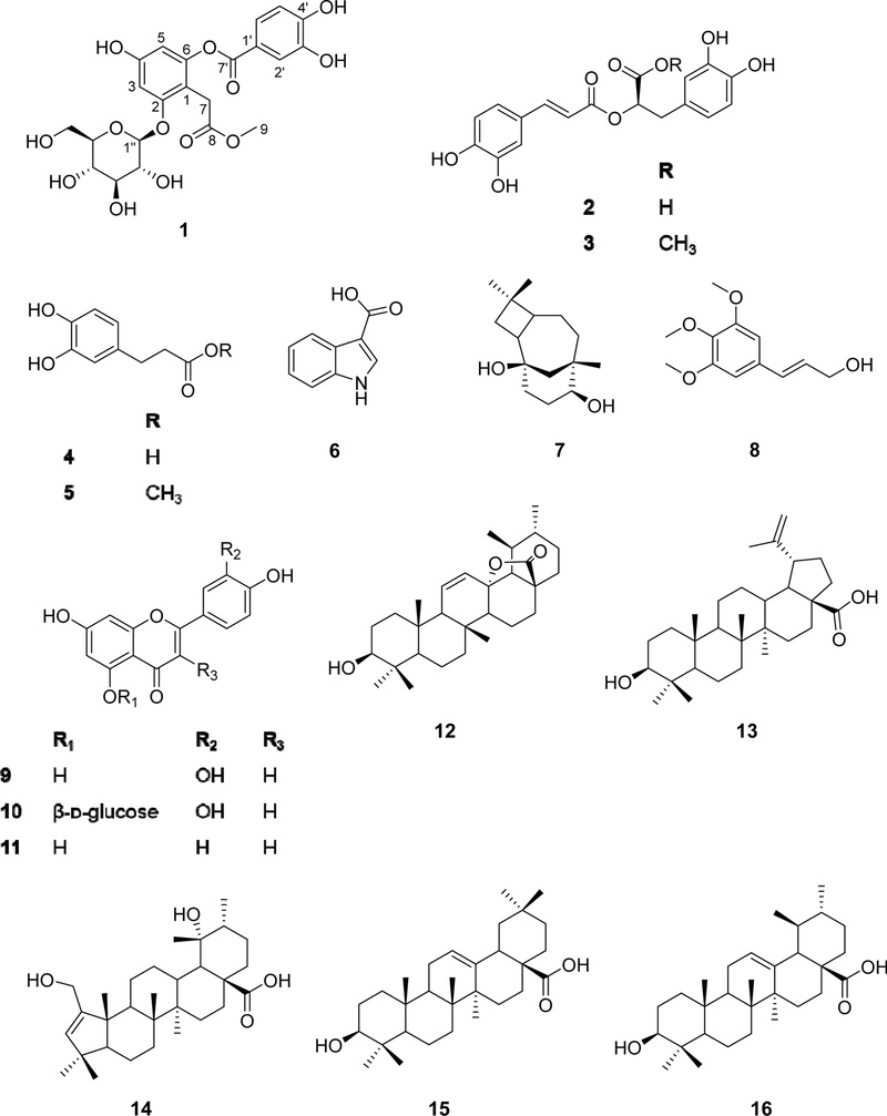
Chemical structures of compounds 1–16.
The 13C-NMR spectrum (Fig. S5) of compound 1 revealed the existence of 22 carbon signals, including two carboxylic carbons at δC 172.6 (C-8) and 165.4 (C-7'). Signals attributable to a β-D-glucopyranosyl unit were observed at δC 103.6 (C-1''), 79.1 (C-5''), 78.8 (C-3''), 75.2 (C-2''), 71.2 (C-4''), and 62.4 (C-6''), along with a methoxy carbon signal at δC 52.0. Comparison of 1H-NMR and 13C-NMR of compound 1 with those of the known depsides, rosarugoside C, showed close similarity, except for the absence of one methoxy group at C-3' in 1.25 The sugar moiety was determined to be attached at C-2 based on the HMBC correlation between the anomeric proton H-1'' (δH 5.49) and C-2 (δC 158.7) (Fig. 2). In addition, the methoxy group was confirmed to C-8 through the HMBC correlation from the OCH3 proton (δH 3.49) to C-8 (δC 172.6). Based on these observations, compound 1 was identified as methyl 6-[(3,4-dihydroxybenzoyloxy)-4-hydroxyphenyl] acetate-2-β-D-glucose. Therefore, the structure of the new depside glucoside (1) was elucidated and named perilloside F.
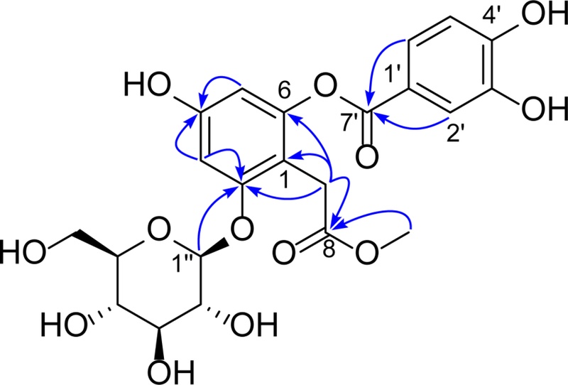
Key HMBC correlation of compound 1.
The inhibitory effects of the 16 compounds isolated from P. frutescens var. acuta were evaluated in RAW 264.7 macrophages by measuring their ability to suppress NO production. As shown in Table 1, compounds 5, 8, 9, and 11 significantly inhibited NO production in RAW 264.7 cells, with IC50 values of 15.35, 17.31, 10.55, and 12.31 μM, respectively. In a previous study, compound 9, luteolin, was reported to be isolated from P. frutescens and showed the NO production inhibition effect with an IC50 value of 6.5 μM in LPS-activated microglia.26 That report is consistent with the findings of this study, further supporting the effects of luteolin and simultaneously confirming the accuracy of the research results. Compounds 13 and 3 also exhibited inhibitory activity, with IC50 values of 39.01 and 65.72 μM, respectively. None of the active compounds showed cytotoxicity at concentrations below 50 μM, indicating that the observed inhibitory effects were not attributable to reduced cell viability.

Inhibition of NO production in RAW 264.7 cells by compounds 1–16.
Compounds 9–11 share the same flavonoid skeleton. Among them, compounds 9 and 11 exhibited potent inhibition of NO production, with IC50 values of 10.55 and 12.31 μM, respectively. In contrast, the glucoside flavonoids, compound 10, did not show inhibitory activity against NO production, suggesting that the 5-O-glucoside substitution results in a decrease in the NO production inhibition effect of flavonoid compounds. The IC50 values presented in Table 1 indicate that glycosylation reduces the inhibitory activity of these flavonoids toward NO production. Compounds 2–5 are phenolic compounds. Of these, compound 3 is an ester derivative of compound 2, while compound 5 is an ester derivative of compound 4. Among these, only compounds 3 and 5 show notable inhibitory activity, with IC50 values of 65.72 and 15.35 μM, respectively. Analysis of their IC50 values suggests that methyl esterification enhances the inhibitory activity of these compounds against NO production.
In summary, P. frutescens var. acuta (Thunb.) Kudo has long been recognized in Asian traditional medicine for its anti-inflammatory properties. In this study, a new depside glucoside, perilloside F (1), along with fifteen known compounds (2–16), was isolated from its aerial parts. Structural elucidation was achieved by spectroscopic methods. Anti-inflammatory activity was evaluated through nitric oxide (NO) inhibition in RAW 264.7 macrophages, where six compounds (3, 5, 8, 9, 11, and 13) showed significant activity. Compound 9 exhibited the strongest inhibition (IC50 = 10.55 μM) without cytotoxicity below 50 μM. Importantly, structure–activity analysis revealed that methyl esterification enhances the anti-inflammatory activity of phenolic acids, whereas 5-O-glycosylation may reduce the activity of flavonoids. These findings not only validate the traditional use of P. frutescens as an anti-inflammatory agent but also provide mechanistic insights into how structural modifications influence bioactivity, highlighting its potential as a valuable source of novel therapeutic leads.
Acknowledgments
This research was supported by the Gyeongsangbuk-do RISE (Regional Innovation System & Education; 2025-RISE-15-107). We thank the Korea Basic Science Institute (KBSI) for mass spectrometric measurements.
Conflicts of Interest
The authors declare no competing financial interests.
References
-
Hou, T.; Netala, V. R.; Zhang, H.; Xing, Y.; Li, H.; Zhang, Z. Molecules 2022, 27, 3578.
[https://doi.org/10.3390/molecules27113578]

-
Ahmed, H. M. Molecules 2018, 24, 102.
[https://doi.org/10.3390/molecules24010102]
-
Huang, B.‐P.; Lin, C.‐H.; Chen, Y.‐C.; Kao, S.‐H. Mol. Med. Rep. 2014, 10, 1077–1083.
[https://doi.org/10.3892/mmr.2014.2298]
-
Simon, L. S. Am. J. Med. 1999, 106, 37S–42S.
[https://doi.org/10.1016/S0002-9343(99)00115-1]
-
Król, M.; Kepinska, M. Int. J. Mol. Sci. 2020, 22, 56.
[https://doi.org/10.3390/ijms22010056]
- Le, T. T.; Ha, M. T.; Han, K.‐H.; Kim, Y.‐B.; Kim, J. A.; Min, B. S. Chem. Biodivers. 2022, 19, e202100986.
-
Woo, E.‐R.; Piao, M. S. Arch. Pharm. Res. 2004, 27, 173–176.
[https://doi.org/10.1007/BF02980102]
-
Sevindik, H. G.; Ozgen, U.; Atila, A.; Er, H. O.; Kazaz, C.; Duman, H. Chem. Pharm. Bull. 2015, 63, 720–725.
[https://doi.org/10.1248/cpb.c14-00877]
-
Putra, N.; Faroliu, G.; Syafni, N.; Nofrizal; Bakhtiar, A.; Arbain, D. J. Math. Fund. Sci. 2021, 53, 231–242.
[https://doi.org/10.5614/j.math.fund.sci.2021.53.2.5]
- Anh, N. T. V.; Le, L. H.; Hang, N. T. M.; Thom, V. T.; Tung, B. T. VNU J. Sci. Med. Pharm. Sci. 2021, 37, 18–23.
-
Thao, V. M.; Thoa, H. T.; Oanh, N. T. T.; Nhiem, N. X.; Hang, N. T. M. VNU J. Sci. Med. Pharm. Sci. 2023, 39, 59–68
[https://doi.org/10.25073/2588-1132/vnumps.4490]
-
Hagemeier, J.; Schneider, B.; Oldham, N. J.; Hahlbrock, K. Proc. Natl. Acad. Sci. U. S. A. 2001, 98, 753–758.
[https://doi.org/10.1073/pnas.98.2.753]
-
Heymann, H.; Tezuka, Y.; Kikuchi, T.; Supriyatna, S. Chem. Pharm. Bull. 1994, 42, 138–146.
[https://doi.org/10.1248/cpb.42.138]
-
Sadik, M. G.; Islam, R.; Rahman, M. M.; Khondkar, P. Fitoterapia 2003, 74, 308–311.
[https://doi.org/10.1016/S0367-326X(03)00041-8]
-
Hung, N. D. H.; Zhao, B. T.; Le, D. D.; Kim, K. Y.; Kim, Y. H.; Yoon, Y. H.; Ko, J. Y.; Woo, K. S.; Woo, M. H. Nat. Prod. Sci. 2016, 22, 140–145.
[https://doi.org/10.20307/nps.2016.22.2.140]
-
Tabaza, Y. Z.; Seong, Z.‐K.; Kim, Y.‐M.; Alshaer, W.; Aburjai, T. A. Trop. J. Pharm. Res. 2024, 23, 77–83.
[https://doi.org/10.4314/tjpr.v23i1.10]
- Fajriah, S.; Megawati, A.; Darmawan, A. J. Trop. Life Sci. 2016, 6, 7–9.
- Ahn, D.; Lee, S. I.; Yang, J. H.; Cho, C. H.; Hwang, Y.‐H.; Park, J.‐H.; Kim, D. K. Nat. Prod. Sci. 2011, 17, 142–146.
-
Topçu, G.; Yapar, G.; Türkmen, Z.; Gören, A. C.; Öksüz, S.; Schilling, J. K.; Kingston, D. G. I. Phytochem. Lett. 2011, 4, 421–425.
[https://doi.org/10.1016/j.phytol.2011.05.002]
-
Bilkisu, A.; Ariyo, A. L.; Umar, A.; Olayemi, A. E.; Isyaka, S. M.; Peter, A. O.; Lanre, O. T. J. Med. Plants Stud. 2024, 12, 48–54.
[https://doi.org/10.22271/plants.2024.v12.i1a.1625]
- Nam, V. V.; Hue, N. T.; Hang, P. T.; Hue, V. T.; Thanh, L. N.; Nhiem, N. X.; Cuong, P. V.; Vuong, N. Q. Vietnam J. Anal. Sci. 2022, 28, 1–6.
-
Taniguchi, S.; Imayoshi, Y.; Kobayashi, E.; Takamatsu, Y.; Ito, H.; Hatano, T.; Sakagami, H.; Tokuda, H.; Nishino, H.; Sugita, D.; Shimura, S.; Yoshida, T. Phytochemistry 2002, 59, 315–323.
[https://doi.org/10.1016/S0031-9422(01)00455-1]
- Maria, F.; Kabouche, Z.; Fujimoto, Y.; Araya, H. Nat. Prod. Commun. 2017, 12, 483–486.
-
Nguyen, T.‐T. A.; Ha, M. T.; Park, S.‐E.; Choi, J. S.; Min, B. S.; Kim, J. A. J. Nat. Prod. 2020, 83, 323–332.
[https://doi.org/10.1021/acs.jnatprod.9b00777]
-
Chang, S. W.; Du, Y. E.; Qi, Y.; Lee, J. S.; Goo, N.; Koo, B. K.; Bae, H. J.; Ryu, J. H.; Jang, D. S. J. Agric. Food Chem. 2019, 67, 7289–7296.
[https://doi.org/10.1021/acs.jafc.9b01228]
-
Kim, J. S.; Lee, H. J.; Lee, M. H.; Kim, J.; Jin, C.; Ryu, J.‐H. Planta Med. 2006, 72, 65–68.
[https://doi.org/10.1055/s-2005-873145]