
Antofine and Related Phenanthroindolizidine Alkaloids in Cancer: Structure–Activity Relationships and Molecular Mechanisms
Abstract
Antofine is a naturally occurring phenanthroindolizidine alkaloid with potent antiproliferative activity against a broad range of cancer cells. Despite its remarkable cellular potency, the therapeutic development of antofine has been limited by poor aqueous solubility, potential toxicity, and an incompletely defined mechanism of action. This review provides an antofine-centered medicinal-chemistry perspective on the structural determinants, pharmacological mechanisms, and translational potential of phenanthroindolizidine alkaloids. Structure–activity relationship studies have identified the C-2 methoxy group, C-13a stereochemistry, tertiary amine, and intact pentacyclic framework as important determinants of biological activity, whereas the C-3 and particularly C-6 positions offer opportunities for property-directed optimization. Polar substitution at C-13a and C-6 has demonstrated that improved solubility, pharmacokinetic exposure, and in vivo efficacy can be achieved while retaining substantial antitumor potency. Mechanistically, antofine modulates multiple cancer-relevant processes, including DNA synthesis, Wnt/β-catenin signaling, P-glycoprotein-associated multidrug resistance, angiogenesis, and Met receptor trafficking. Particularly notable is its ability to promote c-Cbl-dependent Met degradation and disrupt Met endosomal signaling without directly inhibiting Met kinase activity. Collectively, these findings position antofine as a promising scaffold for mechanism-guided anticancer drug development while highlighting the need to further define its direct molecular targets.
Keywords:
Antofine, Phenanthroindolizidine alkaloids, Structure–activity relationship, Anticancer activity, Met endosomal signaling, Multidrug resistanceIntroduction
Natural products have long provided both therapeutic agents and privileged chemical scaffolds for drug discovery. Their value lies not only in biological potency but also in their capacity to access chemical space that is often underrepresented in conventional synthetic libraries. Alkaloids, in particular, have made enduring contributions to oncology, infectious disease therapy, cardiovascular medicine, and neuropharmacology. The phenanthroindolizidine alkaloids represent an unusual and biologically rich alkaloid family whose members are distinguished by a rigid, fused pentacyclic framework comprising a phenanthrene ring system and an indolizidine moiety. Representative members include antofine, tylophorine, tylocrebrine, pergularinine, tylophorinine, and related natural or semisynthetic congeners.1-3
Phenanthroindolizidine alkaloids occur predominantly in plants belonging to the Apocynaceae family, including species of Cynanchum, Tylophora, Vincetoxicum, and Pergularia. Related compounds have also been identified in plants from Moraceae, Lauraceae, Urticaceae, and Acanthaceae, as well as in phytophagous insects that acquire these molecules from host plants. More than one hundred naturally occurring phenanthrene-fused indolizidine alkaloids and related congeners have been reported, illustrating substantial diversity in aromatic substitution, oxidation state, ring fusion, stereochemistry, and peripheral functionalization.4
Antofine is among the best-characterized phenanthroindolizidine alkaloids. It has been isolated from several plant species, including Cynanchum paniculatum, a medicinal plant traditionally used for pain and inflammatory disorders. Antofine possesses a fused phenanthrene–indolizidine skeleton bearing methoxy substituents on the phenanthrene ring and a stereochemically defined ring-junction carbon. The molecule is highly lipophilic, conformationally constrained, and contains a tertiary amine that contributes to its physicochemical and biological behavior. The combination of aromatic surface area, basicity, and a rigid three-dimensional framework appears central to the potent activity of this family.1 The representative structures of antofine and related alkaloids are summarized in Fig. 1.
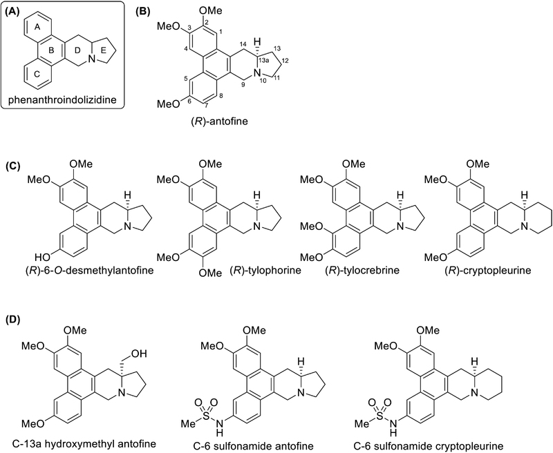
Representative structures of phenanthroindolizidine alkaloids and selected antofine analogues (A) General pentacyclic phenanthroindolizidine scaffold with ring labeling. (B) Chemical structure and atom numbering of natural (R)-antofine. (C) Representative naturally occurring phenanthroindolizidine and related alkaloids, including 6-O-desmethylantofine, tylophorine, tylocrebrine, and cryptopleurine. (D) Selected medicinal-chemistry analogues designed to improve solubility, pharmacokinetic properties, and antitumor activity, including C-13a hydroxymethyl antofine and C-6 sulfonamide derivatives.
The development of antofine as an anticancer lead has benefited from a sequence of complementary studies involving total synthesis, analogue generation, cellular pharmacology, mechanism-of-action analysis, and in vivo efficacy assessment. These studies established several important concepts. First, antofine is not merely a broadly cytotoxic compound; it can inhibit cancer-cell growth through a predominantly cytostatic phenotype in selected settings, with context-dependent effects on cell-cycle progression and apoptotic competence. Second, small structural changes around the phenanthrene ring or at the ring junction can dramatically alter potency, solubility, transporter interaction, and signaling effects. Third, antofine can affect cancer phenotypes that are of high translational importance, including multidrug resistance, receptor tyrosine kinase trafficking, angiogenesis, and growth-factor-dependent survival signaling. Finally, rational analogue design can improve properties such as aqueous solubility and oral bioavailability while preserving meaningful antitumor activity.5
Recent reviews have comprehensively covered the chemistry, natural sources, biosynthesis, biological activities, structural diversity, agricultural applications, toxicity, and synthetic strategies of phenanthroindolizidine alkaloids. In contrast, this review provides an antofine-centered perspective from the standpoint of medicinal chemistry. We focus on the progression from the parent natural product to functionally improved analogues and from descriptive anticancer activity to increasingly refined mechanistic insights.4
Chemical Features and Synthetic Access to Antofine
Structural characteristics of the phenanthroindolizidine scaffold – Antofine belongs to the phenanthroindolizidine class of alkaloids, which is defined by a pentacyclic framework formed through the fusion of a substituted phenanthrene system to an indolizidine ring. This compact scaffold combines several features that are frequently associated with strong biological activity: a rigid polycyclic architecture, a hydrophobic aromatic region, a basic tertiary amine, stereochemical complexity, and a limited number of strategically positioned heteroatom-containing substituents.
The phenanthrene moiety of antofine contains methoxy groups at C-2, C-3, and C-6. These substituents are not merely decorative features inherited from the natural product; they critically influence potency and likely regulate the molecular recognition properties of the scaffold. The fused indolizidine domain contains the tertiary amine and establishes the overall three-dimensional shape of the molecule. The C-13a stereocenter is also a major determinant of biological activity, as shown by comparisons between enantiopure compounds and racemic mixtures.1
The closely related natural products cryptopleurine, tylophorine, and tylocrebrine differ in aromatic substitution pattern and/or ring architecture. Cryptopleurine is generally more potent than antofine in many cancer cell-growth assays, whereas tylophorine and tylocrebrine have been investigated extensively for antiviral, anti-inflammatory, and anticancer activities. Such structural diversity provides an opportunity to define which molecular components are essential for activity and which positions can be modified to improve drug-like properties.1
Importance of asymmetric synthesis – Natural phenanthroindolizidine alkaloids are generally isolated in small quantities, and their complex stereochemistry presents a significant obstacle to broad biological evaluation. Efficient total synthesis is therefore essential not only for supply but also for analogue development. The asymmetric synthesis of R-antofine and R-cryptopleurine established an important foundation for subsequent medicinal chemistry studies. An approach employing stereochemically defined allylic intermediates, an Overman rearrangement, olefin metathesis, and Pictet–Spengler cyclomethylenation enabled preparation of the natural enantiomers of both alkaloids. This synthetic strategy also demonstrated that chirality could be transferred effectively from a chiral building block to the final pentacyclic natural-product scaffold.6
The availability of enantiomerically enriched antofine was particularly important because the biological activity of this class is highly stereodependent. In the original phenanthrene-ring SAR study, natural R-antofine was approximately twofold more potent than racemic antofine against HCT116, HT-1080, and A549 cancer cells. This finding suggested that the cytotoxic activity of the racemate was largely attributable to one enantiomer. Subsequent work with C-13a-substituted analogues further showed that the R-configured compound was markedly more potent than its S-configured counterpart.5,7
Together, these studies demonstrate that stereochemical control is essential for defining meaningful structure–activity relationships within the antofine scaffold and should therefore be incorporated early in analogue design and biological evaluation.
Antofine as a medicinal-chemistry platform – The chemical structure of antofine contains several loci for medicinal-chemistry intervention:
the C-2, C-3, and C-6 substituents on the phenanthrene ring;
the tertiary amine-containing indolizidine ring system;
the C-13a stereocenter and ring-junction geometry; and the E-ring and other positions amenable to polar substitution or scaffold modification.
Not all sites are equally tolerant to modification. Some changes destroy antiproliferative potency, whereas others preserve activity and improve a physicochemical property. The central challenge is therefore to distinguish structural features essential to the pharmacophore from features that can be used to tune solubility, metabolic stability, permeability, and pharmacokinetic properties
Antofine is especially useful as a prototype because its three methoxy substituents allow systematic comparison of positional effects. Furthermore, its potent activity in cancer cells provides a sensitive biological readout for SAR studies. The parent compound, however, also illustrates the limitations of the class. It has low water solubility, and early work suggested that methoxy substitution may contribute to metabolic instability. These limitations motivated the development of polar and bioisosteric modifications at selected positions of the scaffold, particularly C-13a and C-6, to improve physicochemical and pharmacokinetic properties while retaining antiproliferative activity.5, 7, 8
Structure–Activity Relationships of Antofine-Centered Analogues
Phenanthrene-ring substitution as a determinant of potency – A systematic SAR study of antofine demonstrated that the methoxy substituents on the phenanthrene ring are not functionally equivalent. Natural R-antofine showed low-nanomolar growth-inhibitory activity against HCT116, HT-1080, and A549 cells, with IC₅₀ values of approximately 10 nM, whereas racemic antofine was less potent, with IC₅₀ values of approximately 25–30 nM.5
The C-2 position was particularly sensitive to modification. Replacement of the C-2 methoxy group with either an isopropoxy or hydroxy group markedly reduced potency, indicating that bulky substitution and replacement with a hydroxy group are poorly tolerated at this site. Thus, the C-2 methoxy group appears to be an important structural determinant of antiproliferative activity.5 In contrast, the C-3 position was more permissive: isopropoxy and hydroxy substitutions caused only modest changes in potency, suggesting that this site may better tolerate structural diversification.
The C-6 position also proved amenable to optimization. Replacement of the C-6 methoxy group with a hydroxy group yielded 6-O-desmethylantofine, which showed IC₅₀ values close to 1 nM and was more potent than parent antofine. Removal of the C-6 oxygen substituent reduced activity, supporting the importance of an appropriately sized oxygen-containing or hydrogen-bonding substituent at this position.5
Finally, replacement of the 2,3-dimethoxy pattern with a constrained methylenedioxy group caused a marked loss of activity. This finding suggests that the native 2,3-dimethoxy arrangement provides structural features that are not fully reproduced by a rigid methylenedioxy bridge. Overall, the SAR identifies C-2 as highly constrained, C-3 as relatively permissive, and C-6 as a particularly useful site for property-directed analogue design.5 These key structural requirements and modification-tolerant regions are summarized in Fig. 2.
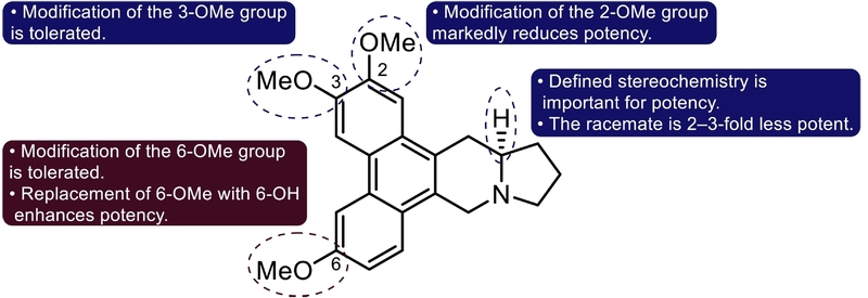
Summary of the structure–activity relationship of antofine at the phenanthrene ring and C-13a stereocenter. The methoxy substituents of antofine exert position-dependent effects on antiproliferative activity. Modification of the C-2 methoxy group markedly reduces potency, indicating that this position is highly constrained. In contrast, structural modification of the C-3 methoxy group is relatively well tolerated. The C-6 methoxy group can also be modified, and replacement with a hydroxy group, as in 6-O-desmethylantofine, enhances antiproliferative potency. In addition, the defined stereochemistry at the C-13a ring junction is important for activity, as racemic antofine is approximately two- to threefold less potent than natural R-antofine. The summary is based on growth-inhibitory activity measured in HCT116, HT-1080, and A549 cancer cells.⁵
C-2 methoxy requirement extends to anti-inflammatory activity – The importance of the C-2 methoxy group was also observed in studies of LPS-induced nitric oxide production in RAW264.7 macrophages. Antofine inhibited LPS-induced nitric oxide production with an IC50 of 2.6 nM under conditions in which activity was measured below overtly cytotoxic concentrations. Several analogues retained activity, but structural changes at C-2 were particularly detrimental. Replacement of the C-2 methoxy group with alternative substituents caused a complete loss of nitric oxide-inhibitory activity, whereas more limited changes at C-3 or C-6 could retain partial activity.9
This SAR pattern closely paralleled that observed for antiproliferative activity, indicating that the C-2 methoxy group represents a common structural requirement for both biological effects. However, this similarity does not establish that the two activities arise from the same direct molecular target.
The importance of chirality at C-13a – C-13a stereochemistry strongly influences the activity of phenanthroindolizidine alkaloids. Natural R-antofine is more potent than the racemate, and the same stereochemical preference was observed with polar analogues. In a study aimed at improving solubility and lowering predicted blood–brain barrier penetration, hydroxymethyl, ester, and carboxylic-acid groups were introduced at C-13a. The ester and carboxylic-acid analogues were essentially inactive at concentrations up to 1 μM, whereas the hydroxymethyl analogue retained submicromolar antiproliferative activity.7
The enantiomeric comparison was particularly informative. The R-configured C-13a hydroxymethyl analogue showed IC₅₀ values of approximately 60–200 nM across a panel of cancer cell lines, whereas the corresponding S-enantiomer was substantially weaker, with IC₅₀ values of approximately 0.5–3.7 μM. Although the R-hydroxymethyl analogue was approximately tenfold less potent than parent antofine, its aqueous solubility was markedly improved, from <1 μg/mL for antofine to 656 μg/mL, and it retained in vivo antitumor activity in A549 and A549-Pa xenograft models.7
These findings establish C-13a stereochemistry as an important determinant of antiproliferative activity and demonstrate that polar modification at this position can improve physicochemical properties without completely sacrificing antitumor activity.
C-6 sulfonamide analogues and oral activity – The C-6 position provided an especially fruitful opportunity for property-directed analogue design. Building on the observation that C-6 hydroxy substitution could enhance activity, methanesulfonamide analogues of antofine and cryptopleurine were synthesized. The C-6 methanesulfonamide analogue of antofine retained low-nanomolar antiproliferative activity, while the corresponding cryptopleurine analogue showed subnanomolar potency across multiple cancer-cell lines, including A549, HCT116, SNU-638, MDA-MB-231, SK-Hep-1, PC-3, and Caki-1 cells.8
The cryptopleurine methanesulfonamide analogue was particularly notable. It was approximately two- to threefold more potent than parent cryptopleurine and showed substantially improved aqueous solubility. In phosphate buffer, the analogue displayed solubility of approximately 6.95 μg/mL compared with <1 μg/mL for cryptopleurine. In a phosphate buffer/ethanol mixture, the solubility improvement was also pronounced. Pharmacokinetic experiments in mice showed lower clearance, a longer apparent oral half-life, increased oral exposure, and oral bioavailability of approximately 30%, compared with approximately 10% for parent cryptopleurine.8
The methanesulfonamide group appears to be a successful C-6 bioisostere because it introduces polarity and a potential hydrogen-bond donor without imposing excessive steric burden. Larger sulfonamide groups, such as isopropyl- or benzenesulfonamide substituents, reduced potency. Acetamide and carbamate analogues also retained activity but were less potent than the methanesulfonamide derivative. These findings emphasize that C-6 is a useful optimization handle, but that substituent size, polarity, hydrogen-bonding properties, and amide acidity must be carefully balanced.8
Antitumor Mechanisms of Antofine
Potent growth inhibition with a context-dependent cytostatic phenotype – Antofine exhibits potent antiproliferative activity in a wide range of human cancer-cell lines. In one study, IC50 values were below 30 nM in colon, lung, breast, gastric, fibrosarcoma, leukemia, and prostate cancer cell lines. HCT116 cells showed an IC50 of approximately 6.3 nM, A549 cells approximately 9.6 nM, and SNU-638 cells approximately 8.9 nM. The activity was preserved in paclitaxel-resistant A549-PA and 5-fluorouracil-resistant HCT116-FU cells, suggesting that antofine can retain growth-inhibitory function in selected chemotherapy-resistant models.10
Despite this striking potency, antofine did not uniformly behave like a conventional apoptosis-inducing cytotoxic agent. In HCT116 cells, treatment with antofine strongly reduced cell proliferation but did not cause a major increase in annexin V- or propidium iodide-positive cells under conditions that produced substantial growth inhibition. These findings support a predominantly cytostatic mechanism at pharmacologically relevant concentrations, rather than direct induction of apoptotic cell death.10
The effects of antofine on cell-cycle distribution varied according to cell type, exposure time, and concentration. In HCT116 cells, low concentrations of antofine produced modest G0/G1 accumulation, whereas higher concentrations or longer exposure could increase the G2/M population. A similar concentration-dependent pattern was observed in A549 cells, while HT-29 cells showed different cell-cycle responses depending on exposure time. Thus, antofine does not appear to induce a single universal cell-cycle phenotype. Instead, it perturbs cell-cycle progression in a context-dependent manner and downregulates several G0/G1-associated regulators, including cyclin D1, cyclin E, and CDK4.10
DNA synthesis inhibition and Wnt/β-catenin modulation – Antofine inhibited DNA synthesis in HCT116 cells, as demonstrated by reduced BrdU incorporation. Earlier studies on phenanthroindolizidine alkaloids have implicated suppression of DNA and protein synthesis, inhibition of dihydrofolate reductase, and interactions with bulged DNA structures. However, these observations do not establish the direct molecular target responsible for the DNA synthesis inhibition induced by antofine in HCT116 cells.10
In HCT116 cells, antofine also reduced β-catenin/Tcf transcriptional activity in a TOPflash reporter assay. In both HCT116 and SW480 colorectal cancer cells, antofine reduced β-catenin and cyclin D1 protein expression. Because cyclin D1 is a canonical downstream target of Wnt/β-catenin signaling and an important regulator of G1 progression, suppression of this pathway may contribute to the growth-inhibitory effects of antofine in colorectal cancer cells.10
The mechanistic relationship between DNA synthesis inhibition and Wnt/β-catenin suppression remains unclear. It is not yet known whether these effects reflect direct pathway engagement by antofine or secondary consequences of broader changes in cellular biosynthetic or transcriptional processes. Further studies are therefore needed to distinguish primary molecular targets from downstream signaling effects.
Overcoming P-glycoprotein-associated multidrug resistance – Multidrug resistance is a major barrier to effective chemotherapy, and P-glycoprotein (P-gp), encoded by MDR1/ABCB1, contributes to resistance by reducing intracellular drug accumulation. In paclitaxel-resistant A549-PA cells, which were more than 200-fold resistant to paclitaxel, antofine retained potent antiproliferative activity with an IC₅₀ of approximately 18 nM, similar to that in parental A549 cells (~16 nM). Antofine reduced P-gp protein and MDR1 mRNA expression, increased intracellular rhodamine 123 accumulation, and synergistically enhanced paclitaxel-mediated growth inhibition.11,12
These findings suggest that antofine can counter P-gp-associated resistance through both downregulation of P-gp expression and suppression of its efflux function. A similar phenotype was retained in the more water-soluble C-13a hydroxymethyl analogue, which remained active in A549-PA cells, suppressed P-gp/MDR1, enhanced rhodamine 123 accumulation, and reduced tumor growth and P-gp expression in A549-PA xenografts.7
Met degradation and endosomal signaling inhibition – The most distinctive mechanistic advance in the pharmacology of antofine is the identification of negative regulation of Met endosomal signaling. Met, also known as hepatocyte growth factor receptor, is a receptor tyrosine kinase involved in cell proliferation, survival, motility, invasion, epithelial–mesenchymal transition, and metastasis. Aberrant Met activation may arise through amplification, mutation, overexpression, ligand-dependent stimulation, or dysregulated receptor trafficking. Although small-molecule Met kinase inhibitors can be effective in selected settings, kinase-domain mutations and alternative signaling mechanisms can limit their clinical activity.13-16
Antofine has been shown to negatively regulate Met endosomal signaling, providing a distinct mechanism from conventional Met kinase inhibition. In Caki-1 renal cancer cells, which harbor a V1238I Met kinase-domain mutation and exhibit high Met activation, antofine potently suppressed phospho-Met and cell proliferation despite resistance to the Met inhibitors cabozantinib and BMS777607. Importantly, antofine did not directly inhibit Met kinase activity in an enzyme assay.17
Instead, antofine promoted c-Cbl-dependent proteasomal degradation of Met. It increased Met ubiquitination and Met–c-Cbl association, while Met degradation was blocked by MG132 but not chloroquine. Antofine also enhanced degradation of cell-surface Met and inhibited Met endocytosis and recycling, thereby disrupting the constitutive shuttling of mutant Met between the plasma membrane and endosomal compartments.17 A proposed model for antofine-mediated disruption of Met trafficking and endosomal signaling is shown in Fig. 3.
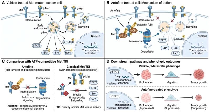
Proposed mechanism by which antofine disrupts Met trafficking and endosomal signaling in Met-mutant cancer cells. (A) In vehicle-treated Met-mutant cancer cells, activated Met undergoes internalization into early endosomal compartments and subsequent recycling to the plasma membrane, thereby sustaining Met-associated signaling through STAT3, ERK, Src, and Rac1 and promoting transcriptional activation. (B) Antofine enhances c-Cbl association with Met, increases Met ubiquitination, and promotes proteasome-dependent receptor degradation while reducing Met internalization and recycling. These effects attenuate downstream signaling, including STAT3 phosphorylation and nuclear signaling. (C) Antofine acts through a mechanism distinct from classical ATP-competitive Met tyrosine kinase inhibitors. Whereas conventional Met TKIs directly block kinase activity, antofine primarily modulates receptor turnover and trafficking, thereby suppressing endosomal signaling. (D) The resulting attenuation of Met-associated signaling is linked to reduced proliferative and migratory phenotypes and suppressed tumor growth. The schematic summarizes the proposed mechanistic model based on cellular and in vivo observations.
This disruption attenuated downstream Met signaling, reducing Grb2 and Rac1 levels, phosphorylation of Src, ERK1/2, and STAT3, Met–STAT3 association, and nuclear accumulation of STAT3. Antofine also inhibited Caki-1 cell migration. In a Caki-1 xenograft model, oral antofine inhibited tumor growth by approximately 59% and reduced Met and STAT3 signaling in tumor tissues.17
Together, these findings identify Met endosomal signaling as a potential mechanism underlying the antitumor activity of antofine and suggest a therapeutic strategy for Met-mutated or Met TKI-resistant cancers.
Anti-angiogenic activity – Antofine also exhibits anti-angiogenic activity in endothelial models. In VEGF-stimulated HUVECs, antofine inhibited proliferation with an IC₅₀ of approximately 36 nM and suppressed endothelial migration and capillary-like tube formation. It also inhibited vascular sprouting in mouse embryonic stem cell-derived endothelial cultures and ex vivo mouse aortic ring assays.18
Mechanistically, antofine suppressed VEGF-induced VEGFR2 phosphorylation and downstream AKT/mTOR/p70S6K and ERK signaling, while promoting AMPK activation and reducing HIF-1α expression. It also decreased c-Myc, cyclin D1, and CDK4 expression and increased p53. These findings suggest that antofine interferes with multiple processes required for angiogenesis, including endothelial proliferation, migration, and vascular structure formation.18
Summary of the antitumor activities and mechanisms of antofine – Taken together, the available studies indicate that antofine exerts broad antitumor effects through multiple cellular processes, including inhibition of proliferation and DNA synthesis, modulation of Wnt/β-catenin signaling, suppression of P-glycoprotein-associated drug resistance, disruption of Met endosomal signaling, and inhibition of angiogenesis. These effects extend beyond direct cancer-cell growth inhibition and include modulation of both tumor-cell signaling and the vascular compartment. The major pharmacological actions of antofine are illustrated in Fig. 4.
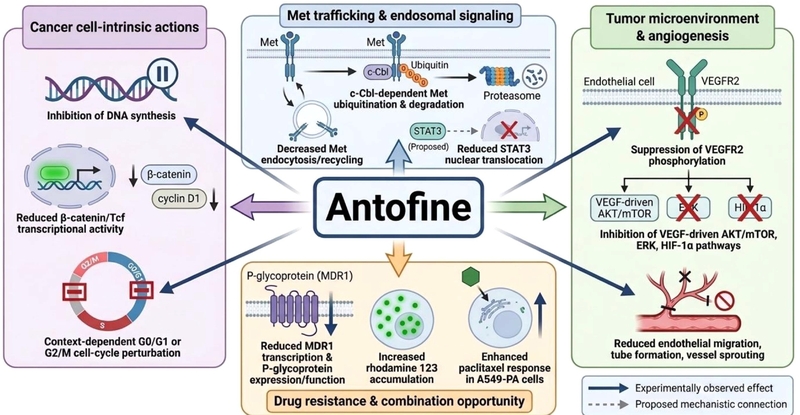
Overview of the major antitumor mechanisms and pharmacological actions of antofine. Antofine exerts pleiotropic anticancer effects across multiple cellular compartments and signaling contexts. In cancer cells, antofine inhibits DNA synthesis, suppresses β-catenin/Tcf transcriptional activity and cyclin D1 expression, and induces context-dependent perturbation of cell-cycle progression. Antofine also modulates Met receptor trafficking by promoting c-Cbl-dependent ubiquitination and proteasomal degradation of Met, while reducing receptor endocytosis/recycling and attenuating downstream STAT3 signaling. In drug-resistant cancer cells, antofine decreases MDR1/P-glycoprotein expression and function, increases intracellular rhodamine 123 accumulation, and enhances the response to paclitaxel. In the tumor microenvironment, antofine suppresses VEGFR2 phosphorylation and downstream VEGF-driven AKT/mTOR, ERK, and HIF-1α signaling, resulting in reduced endothelial-cell migration, tube formation, and vessel sprouting. Solid arrows indicate experimentally observed effects, whereas dashed arrows denote proposed mechanistic connections.
However, the pleiotropic pharmacology of antofine should not be interpreted as evidence that each affected pathway represents an independent primary molecular target. At least some of these phenotypes may arise as downstream consequences of one or more upstream molecular interactions. Identification of the direct molecular targets of antofine will therefore be essential for distinguishing primary mechanisms from downstream effects and for guiding its rational development as an anticancer agent.
Antofine exemplifies both the promise and the challenges of natural-product-based anticancer drug discovery. Its rigid phenanthroindolizidine scaffold supports potent antiproliferative activity, but limited solubility and other drug-like liabilities have complicated its therapeutic development. Progress has therefore relied on integrating structure–activity studies, synthetic modification, mechanistic pharmacology, and in vivo evaluation.
SAR studies have identified several structural features that are important for biological activity, including the C-2 methoxy group, C-13a stereochemistry, the tertiary amine, and the intact pentacyclic framework. By contrast, the C-3 and particularly the C-6 positions provide useful opportunities for optimization. Polar modification at C-13a and methanesulfonamide substitution at C-6 demonstrate that physicochemical and pharmacokinetic properties can be improved while retaining substantial antitumor activity.
Mechanistically, antofine displays pleiotropic antitumor effects involving inhibition of DNA synthesis, modulation of Wnt/β-catenin signaling, suppression of P-glycoprotein-associated drug resistance, inhibition of angiogenesis, and negative regulation of Met endosomal signaling. Among these, the promotion of c-Cbl-dependent Met degradation together with inhibition of Met endocytosis and recycling provides a particularly distinctive mechanism that expands the pharmacology of phenanthroindolizidine alkaloids beyond conventional cytotoxicity.
Future development will require identification of the direct molecular targets of antofine, clarification of the hierarchy among its multiple cellular effects, selection of responsive tumor contexts, and further optimization of pharmacokinetic properties and safety. Such efforts may enable antofine and its analogues to serve as mechanism-guided leads for the development of anticancer agents, particularly in treatment-resistant or signaling-dependent tumors.
Acknowledgments
This research was supported by the National Research Foundation of Korea (NRF) grant funded by the Korean government (Grant No. RS-2026-25474641).
Conflicts of Interest
The author declares no conflict of interest.
References
-
de Fatima Pereira, M.; Rochais, C.; Dallemagne, P. Anticancer Agents Med. Chem. 2015, 15, 1080–1091.
[https://doi.org/10.2174/1871520615666150520143600]

-
Mostafa, E. M.; Musa, A.; Mohammed, H. A.; Alzarea, A. I.; Abdelgawad, M. A.; Al-Sanea, M. M.; Ismail, A.; Zafar, A.; Elmowafy, M.; Selim, S.; Khan, R. A. Plants (Basel) 2023, 12.
[https://doi.org/10.3390/plants12051143]
-
Jia, X. H.; Zhao, H. X.; Du, C. L.; Tang, W. Z.; Wang, X. J. Phytochem. Rev. 2021, 20, 845–868.
[https://doi.org/10.1007/s11101-020-09723-3]
-
Li, X.; Wang, Y.; Chang, X.; Li, X.; Song, J.; Kuang, H.; Yang, L.; Jiang, H. Eur. J. Med. Chem. 2026, 319, 119265.
[https://doi.org/10.1016/j.ejmech.2026.119265]
-
Fu, Y.; Lee, S. K.; Min, H. Y.; Lee, T.; Lee, J.; Cheng, M.; Kim, S. Bioorg. Med. Chem. Lett. 2007, 17, 97–100.
[https://doi.org/10.1016/j.bmcl.2006.09.080]
-
Kim, S.; Lee, T.; Lee, E.; Lee, J.; Fan, G. J.; Lee, S. K.; Kim, D. J. Org. Chem. 2004, 69, 3144–3149.
[https://doi.org/10.1021/jo049820a]
-
Kwon, Y.; Song, J.; Lee, B.; In, J.; Song, H.; Chung, H. J.; Lee, S. K.; Kim, S. Bioorg. Med. Chem. 2013, 21, 1006–1017.
[https://doi.org/10.1016/j.bmc.2012.11.039]
-
Kwon, Y.; Song, J.; Lee, H.; Kim, E. Y.; Lee, K.; Lee, S. K.; Kim, S. J. Med. Chem. 2015, 58, 7749–7762.
[https://doi.org/10.1021/acs.jmedchem.5b00764]
-
Min, H. Y.; Song, S. H.; Lee, B.; Kim, S.; Lee, S. K. Chem. Biodivers. 2010, 7, 409–414.
[https://doi.org/10.1002/cbdv.200900040]
-
Min, H. Y.; Chung, H. J.; Kim, E. H.; Kim, S.; Park, E. J.; Lee, S. K. Biochem. Pharmacol. 2010, 80, 1356–1364.
[https://doi.org/10.1016/j.bcp.2010.07.026]
-
Ling, V. Cancer Chemother. Pharmacol. 1997, 40, S3–8.
[https://doi.org/10.1007/s002800051053]
-
Kim, E. H.; Min, H. Y.; Chung, H. J.; Song, J.; Park, H. J.; Kim, S.; Lee, S. K. Food Chem. Toxicol. 2012, 50, 1060–1065.
[https://doi.org/10.1016/j.fct.2011.11.008]
-
Birchmeier, C.; Birchmeier, W.; Gherardi, E.; Vande Woude, G. F. Nat. Rev. Mol. Cell Biol. 2003, 4, 915–925.
[https://doi.org/10.1038/nrm1261]
-
Gherardi, E.; Birchmeier, W.; Birchmeier, C.; Vande Woude, G. Nat. Rev. Cancer 2012, 12, 89–103.
[https://doi.org/10.1038/nrc3205]
-
Joffre, C.; Barrow, R.; Ménard, L.; Calleja, V.; Hart, I. R.; Kermorgant, S. Nat. Cell Biol. 2011, 13, 827–837.
[https://doi.org/10.1038/ncb2257]
-
Tiedt, R.; Degenkolbe, E.; Furet, P.; Appleton, B. A.; Wagner, S.; Schoepfer, J.; Buck, E.; Ruddy, D. A.; Monahan, J. E.; Jones, M. D.; et al. Cancer Res. 2011, 71, 5255–5264.
[https://doi.org/10.1158/0008-5472.CAN-10-4433]
-
Song, J.; Kwon, Y.; Kim, S.; Lee, S. K. Chem. Biol. 2015, 22, 504–515.
[https://doi.org/10.1016/j.chembiol.2015.03.011]
-
Oh, J.; Kim, G. D.; Kim, S.; Lee, S. K. Food Chem. Toxicol. 2017, 107, 201–207.
[https://doi.org/10.1016/j.fct.2017.06.036]