
Inhibitory Effects of Extracts of Ailanthus altissima var. altissima and Lycianthes beckneriana on Wnt/β-Catenin Signaling in Human Colorectal Cancer Cells
Abstract
The canonical Wnt/β-catenin signaling pathway is aberrantly activated in most colorectal cancers and is therefore regarded as an attractive molecular target for the discovery of anticancer agents from natural products. In the present study, the effects of the 95% ethanol extracts of Ailanthus altissima var. altissima (AAE) and Lycianthes beckneriana D’Arcy (LBE) on the growth and the Wnt/β-catenin signaling of human colorectal cancer cells were investigated. Both extracts inhibited the proliferation of HCT116 cells in a concentration-dependent manner after 72 h of treatment, with IC50 values of 9.32 and 6.27 μg/mL, respectively. In HEK293 cells in which β-catenin/T-cell factor (TCF) transcription was ectopically activated by cotransfection with the TOPflash reporter and the TCF4 and β-catenin expression vectors, treatment with AAE or LBE for 24 h suppressed the reporter activity, with IC50 values of 13.80 and 12.92 μg/mL, respectively. Both extracts also decreased the TOPflash activity in HCT116 cells, in which Wnt/β-catenin signaling is constitutively activated, with IC50 values of 3.26 and 12.67 μg/mL, respectively. Consistent with the reporter gene data, the mRNA levels of the Wnt target genes CMYC, CCND1, and BIRC5 were down-regulated after 12 h of treatment, and the levels of the corresponding c-Myc, cyclin D1, and survivin proteins were reduced after 24 h. These findings indicate that the growth-inhibitory activity of AAE and LBE in colorectal cancer cells is associated with the suppression of β-catenin/TCF-mediated transcription and suggest that both extracts are potential sources of Wnt/β-catenin signaling inhibitors.
Keywords:
Ailanthus altissima var. altissima, Lycianthes beckneriana D’Arcy, Colorectal cancer, Wnt/β-catenin signaling, Human colorectal cancerIntroduction
Colorectal cancer (CRC) is one of the most frequently diagnosed malignancies and a leading cause of cancer-related death worldwide.1 Although surgery, cytotoxic chemotherapy, molecularly targeted agents, and immunotherapy have improved the clinical outcome of patients with CRC, recurrence and acquired resistance remain common in advanced disease, and anticancer agents that act through distinct molecular mechanisms are still required.2
The canonical Wnt/β-catenin signaling pathway governs cell proliferation, differentiation, tissue homeostasis, and stem cell maintenance.3,4 In the absence of Wnt ligands, cytosolic β-catenin is recruited into a destruction complex composed of adenomatous polyposis coli (APC), Axin, glycogen synthase kinase 3β, and casein kinase 1α, and is sequentially phosphorylated, ubiquitinated, and degraded by the proteasome. The binding of Wnt ligands to the Frizzled receptor and its coreceptor, low-density lipoprotein receptor-related protein 5/6, dissociates this complex and allows β-catenin to accumulate and translocate into the nucleus, where it associates with T-cell factor/lymphoid enhancer-binding factor (TCF/LEF) transcription factors and induces the transcription of Wnt target genes.5
Aberrant activation of this pathway is a hallmark of colorectal tumorigenesis. Loss-of-function mutations in APC and activating mutations in CTNNB1 stabilize β-catenin and sustain β-catenin/TCF-dependent transcription in colorectal cancer cells.6 Among the best characterized Wnt target genes are CMYC, CCND1, and BIRC5, which encode c-Myc, cyclin D1, and survivin, respectively.7–9 These proteins promote cell-cycle progression and cell survival and thereby link constitutive Wnt signaling to the growth of colorectal tumors. The identification of inhibitors of β-catenin/TCF-responsive transcription is therefore considered an attractive strategy for the development of anticancer agents against CRC.
Natural products have long served as a productive source of Wnt signaling modulators. Using the β-catenin/TCF-responsive TOPflash reporter gene assay as a screening system, plant extracts and their constituents, including 2-hydroxycinnamaldehyde, periplocin, and esculetin, have been shown to suppress β-catenin/TCF transcriptional activity and the expression of Wnt target genes, together with the inhibition of colorectal cancer cell growth.10–12 These findings indicate that the TOPflash reporter gene assay is a practical screening system for identifying natural sources of Wnt pathway inhibitors.
Ailanthus altissima (Mill.) Swingle (Simaroubaceae) is a chemically rich species that accumulates quassinoids, alkaloids, phenylpropanoids, and triterpenoids, and its bark has long been used in traditional medicine in East Asia.13 Ailanthone, the representative quassinoid of this plant, exhibits antitumor activity in a variety of cancer models,14 including human colorectal cancer cells, in which it has been reported to act through the suppression of STAT3 signaling.15 The effect of A. altissima on β-catenin/TCF-mediated transcription in colorectal cancer cells, however, has not been examined. Lycianthes beckneriana D’Arcy (Solanaceae) is a Neotropical species for which no biological activity has been reported to date.
Herein, the 95% ethanol extracts of A. altissima var. altissima (AAE) and L. beckneriana (LBE) were examined for their growth-inhibitory activity in HCT116 human colorectal cancer cells and for their effects on Wnt/β-catenin signaling. β-Catenin/TCF-responsive transcription was evaluated in an ectopically activated HEK293 system and in HCT116 cells harboring constitutively active Wnt signaling, and the expression of Wnt target genes was subsequently determined at the mRNA and protein levels.
Experimental
General experimental procedures – Dulbecco’s modified Eagle’s medium (DMEM), Roswell Park Memorial Institute 1640 medium (RPMI 1640), fetal bovine serum (FBS), antibiotic-antimycotic solution, TRI reagent, and Lipofectamine 2000 were purchased from Invitrogen (Grand Island, NY, USA). Sulforhodamine B (SRB), trichloroacetic acid (TCA), and dimethyl sulfoxide (DMSO) were obtained from Sigma-Aldrich (St. Louis, MO, USA). The pTOPflash (pGL3-OT), pFOPflash (pGL3-OF), and Renilla reporter plasmids were obtained from Upstate Biotechnology (Lake Placid, NY, USA). The TCF4 and pcDNA β-catenin expression vectors were those described previously.10–12 The Dual-Luciferase Reporter Assay System and the Reverse Transcription System were purchased from Promega (Madison, WI, USA), and iQ SYBR Green Supermix was obtained from Bio-Rad (Hercules, CA, USA). Antibodies against c-Myc and cyclin D1 were purchased from BD Biosciences (San Diego, CA, USA), and antibodies against survivin and β-actin were purchased from Santa Cruz Biotechnology (Dallas, TX, USA).
Plant materials and extracts – Two 95% ethanol plant extracts were used in the present study. Ailanthus altissima var. altissima (Simaroubaceae), whose local name is chouchun, was collected in China and was registered under the barcode PBCN12837, and Lycianthes beckneriana D’Arcy (Solanaceae) was collected in Costa Rica and was registered under the barcode PBCR10601. For both plants, the leaves and stems were extracted with 95% ethanol, and the yields of the dried extracts were 19.72 ± 0.19 mg and 24.46 ± 0.15 mg, respectively (Table 1). Each extract was dissolved in DMSO to prepare a stock solution and was diluted with culture medium immediately before treatment. Control cells received an equal volume of the vehicle alone.

Information on the plant materials from which the 95% ethanol extracts were prepared.
Cell culture – HCT116 human colorectal cancer cells and HEK293 human embryonic kidney cells were purchased from the American Type Culture Collection (Manassas, VA, USA). HEK293 cells were maintained in DMEM and HCT116 cells in RPMI 1640, each supplemented with 10% FBS and antibiotic-antimycotic solution (100 units/mL penicillin G sodium, 100 μg/mL streptomycin, and 250 ng/mL amphotericin B). The cells were cultured at 37°C in a humidified atmosphere containing 5% CO2.
Cell proliferation assay – The growth-inhibitory activity of the extracts was determined by the SRB assay as described previously.12,16 HCT116 cells were seeded in 96-well plates (7 × 10³ cells/well) and were incubated for 30 min (for day-zero controls) or treated with the indicated concentrations of AAE or LBE for 72 h. After incubation, the cells were fixed with 10% TCA, dried, and stained with 0.4% SRB in 1% acetic acid. The unbound dye was washed out with 1% acetic acid, and the protein-bound dye was dissolved in 10 mM Tris (pH 10.0). The absorbance was measured at 515 nm, and cell viability was expressed as a percentage of that of the vehicle-treated control. IC50 values were calculated by nonlinear regression analysis using TableCurve 2D v5.01 software (Systat Software Inc., Richmond, CA, USA).
Transfection and luciferase reporter gene assay – Transient transfections were performed using Lipofectamine 2000 as described previously.10–12 Cells were seeded in 48-well plates at a density of 1.5 × 10⁴ cells/well. After 24 h of incubation, the cells were transfected with 0.1 μg of the TOPflash or FOPflash reporter plasmid and 0.005 μg of the Renilla luciferase vector for normalization. To activate β-catenin/TCF-responsive transcription ectopically, HEK293 cells were additionally cotransfected with 0.02 μg of the pcDNA β-catenin expression vector and 0.004 μg of the TCF4 expression vector. At 24 h after transfection, the cells were treated with AAE or LBE at the indicated concentrations for an additional 24 h, and the cell lysates were subjected to a dual luciferase activity assay using the Dual-Luciferase Reporter Assay System according to the manufacturer’s instructions. Firefly luciferase activity was normalized to Renilla activity and is expressed relative to that of the vehicle-treated control. All experiments were performed in triplicate.
RNA extraction and real-time PCR – HCT116 cells were treated with the indicated concentrations of AAE or LBE for 12 h. Total RNA was extracted using TRI reagent and was reverse-transcribed using the Reverse Transcription System according to the manufacturer’s instructions. Real-time PCR was performed using iQ SYBR Green Supermix. The relative expression levels of CMYC, CCND1, and BIRC5 were normalized to those of ACTB and were calculated by the comparative CT (2−ΔΔCT) method.17 The sequences of the primers used are listed in Table 2.
Sequences of the target gene-specific primers used for real-time PCR.
Western blot analysis – HCT116 cells were treated with the indicated concentrations of AAE or LBE for 24 h. Total cell lysates were prepared in 2× sample loading buffer containing 250 mM Tris-HCl (pH 6.8), 4% sodium dodecyl sulfate (SDS), 10% glycerol, 0.006% bromophenol blue, 2% β-mercaptoethanol, 50 mM sodium fluoride, and 5 mM sodium orthovanadate. Protein concentrations were determined by the bicinchoninic acid method. Equal amounts of protein (5–20 μg) were resolved by 8–12% SDS-polyacrylamide gel electrophoresis and were transferred to polyvinylidene fluoride membranes. The membranes were blocked with 5% bovine serum albumin in phosphate-buffered saline containing 0.1% Tween-20 and were then probed overnight at 4°C with antibodies against c-Myc, cyclin D1, survivin, or β-actin. After incubation with the appropriate horseradish peroxidase-conjugated secondary antibodies, the protein bands were visualized using an enhanced chemiluminescence detection kit. β-Actin was used as an internal standard. For quantitative analysis, the band intensities were measured by densitometry using ImageJ software (National Institutes of Health, Bethesda, MD, USA), normalized to those of β-actin, and expressed relative to those of the vehicletreated control. Densitometric data were obtained from three independent experiments.
Statistical analysis – The data are presented as the mean values ± SD for the indicated number of independently performed experiments and are representative of the results of at least three independent experiments. The significance of the differences between data was assessed using Student’s t-test or a one-way analysis of variance (ANOVA) coupled with Dunnett’s t-test. Values of p < 0.05 were considered statistically significant.
Results and Discussion
The growth-inhibitory activities of AAE and LBE were first evaluated in HCT116 human colorectal cancer cells using the SRB assay. Both extracts reduced cell viability in a concentration-dependent manner after 72 h of treatment (Fig. 1). At 1.6, 8, and 40 μg/mL, AAE decreased the cell viability to 90.8%, 55.0%, and 5.4% of that of the vehicle-treated control, respectively, with an IC50 value of 9.32 μg/mL (Fig. 1A). LBE was somewhat more potent, reducing the cell viability to 70.0%, 31.2%, and 11.7% at the same concentrations, with an IC50 value of 6.27 μg/mL (Fig. 1B). 95% ethanol extracts thus exhibited marked antiproliferative activity against HCT116 cells.
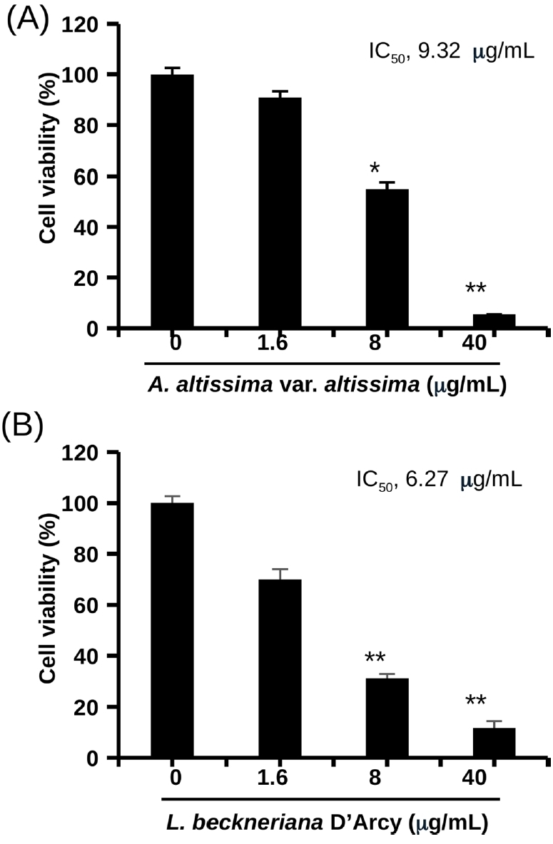
Growth-inhibitory effects of the 95% ethanol extracts of Ailanthus altissima var. altissima (AAE) and Lycianthes beckneriana D’Arcy (LBE) in HCT116 human colorectal cancer cells. HCT116 cells were treated with the indicated concentrations of (A) AAE or (B) LBE for 72 h, and cell viability was determined by the SRB assay. IC50 values were calculated by nonlinear regression analysis. The data are expressed as the mean values ± SD (n = 3) and are representative of three separate determinations. *p < 0.05 and **p < 0.01 compared with the vehicle-treated control.
Because the growth of colorectal cancer cells is largely dependent on constitutively active Wnt/β-catenin signaling, the effects of the extracts on β-catenin/TCF-responsive transcription were examined next. In HEK293 cells, cotransfection of the TOPflash reporter with the TCF4 and pcDNA β-catenin expression vectors markedly increased the luciferase activity compared with that of cells transfected with TOPflash alone, which indicates that the pathway was successfully activated. Treatment with AAE or LBE for 24 h suppressed this activated reporter signal in a concentration-dependent manner (Fig. 2). AAE inhibited the reporter activity with an IC50 value of 13.80 μg/mL (Fig. 2A), and LBE showed a comparable potency, with an IC50 value of 12.92 μg/mL (Fig. 2B). These results indicate that both extracts contain constituents that interfere with the transcriptional output of the canonical Wnt pathway.
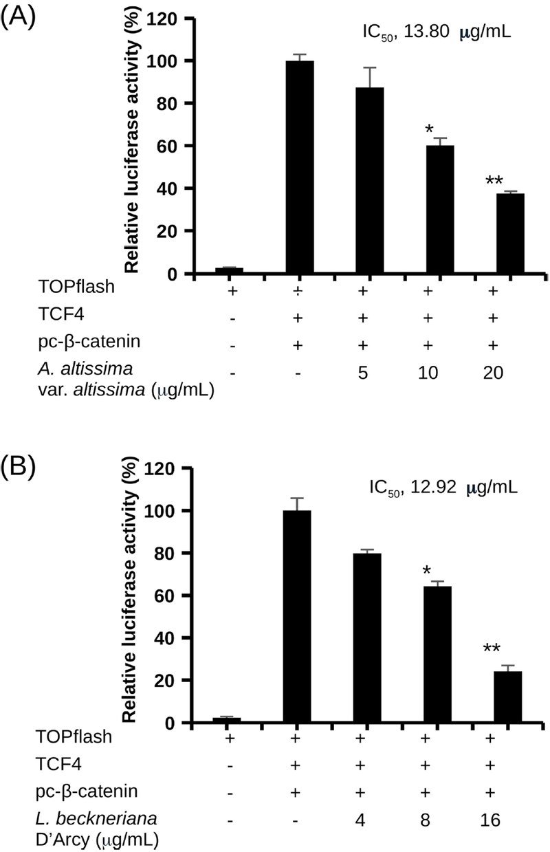
Inhibitory effects of AAE and LBE on β-catenin/TCF-responsive transcriptional activity in HEK293 cells. HEK293 cells were cotransfected with the TOPflash and Renilla reporter gene plasmids and the TCF4 and pc-β-catenin expression vectors for 24 h, and were subsequently treated with the indicated concentrations of (A) AAE or (B) LBE for an additional 24 h. Firefly luciferase activity was normalized to Renilla activity and is expressed relative to that of the activated vehicle-treated control. The data are expressed as the mean values ± SD (n = 3) and are representative of three separate determinations. *p < 0.05 and **p < 0.01 compared with the activated control.
The extracts were then evaluated in HCT116 cells, in which Wnt/β-catenin signaling is constitutively activated by a heterozygous deletion of codon 45 of the β-catenin gene that prevents the phosphorylation and degradation of the protein.6 As expected, the TOPflash activity of these cells was substantially higher than that of the mutant FOPflash reporter, which was used as a negative control. A 24 h treatment with AAE reduced the endogenous TOPflash activity in a concentration-dependent manner, with an IC50 value of 3.26 μg/mL (Fig. 3A), whereas LBE suppressed the same reporter with an IC50 value of 12.67 μg/mL (Fig. 3B). The inhibitory effects observed in the reconstituted HEK293 system were therefore reproduced in a colorectal cancer cell line carrying constitutively active β-catenin signaling. The IC50 value of AAE in HCT116 cells was lower than that in the ectopically activated HEK293 system. However, because the two reporter systems differ in cell type, in the mode of pathway activation (overexpression of β-catenin and TCF4 versus an endogenous activating CTNNB1 mutation), and in the transfection conditions, the IC50 values obtained in the two systems are not directly comparable, and this difference does not by itself indicate that AAE acts preferentially on the endogenously activated pathway.
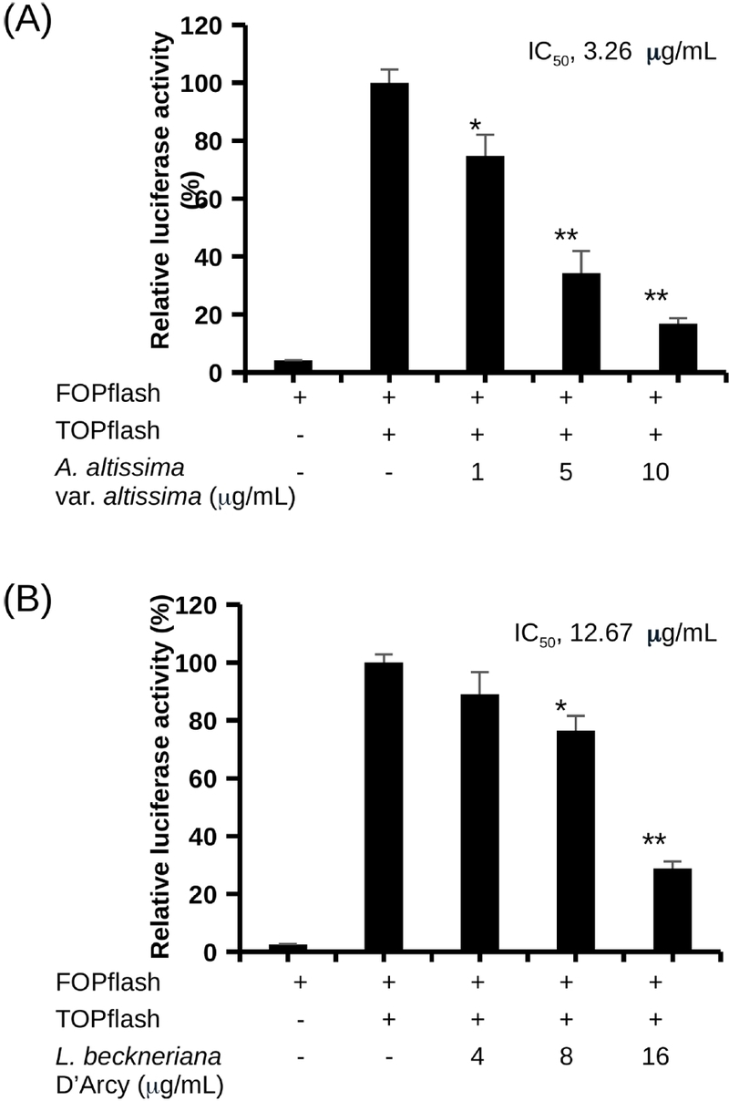
Suppression of endogenous β-catenin/TCF transcriptional activity by AAE and LBE in HCT116 cells. HCT116 cells were transfected with the TOPflash or FOPflash plasmid together with the Renilla reporter gene plasmid for 24 h, and were subsequently treated with the indicated concentrations of (A) AAE or (B) LBE for an additional 24 h. Luciferase activity was normalized to Renilla activity and is expressed relative to that of the TOPflash-transfected vehicle-treated control. The data are expressed as the mean values ± SD (n = 3) and are representative of three separate determinations. *p < 0.05 and **p < 0.01 compared with the TOPflash control.
To determine whether the inhibition of the reporter activity was accompanied by the suppression of endogenous Wnt-regulated genes, HCT116 cells were treated with the extracts for 12 h and the mRNA levels of CMYC, CCND1, and BIRC5 were measured by real-time PCR (Fig. 4). All three transcripts were down-regulated by both extracts in a concentration-dependent manner. At 10 μg/mL, AAE reduced the relative levels of CMYC, CCND1, and BIRC5 to approximately 0.35, 0.38, and 0.31 of those of the vehicle-treated control, respectively (Fig. 4A), and at 16 μg/mL, LBE reduced them to approximately 0.44, 0.58, and 0.60, respectively (Fig. 4B).
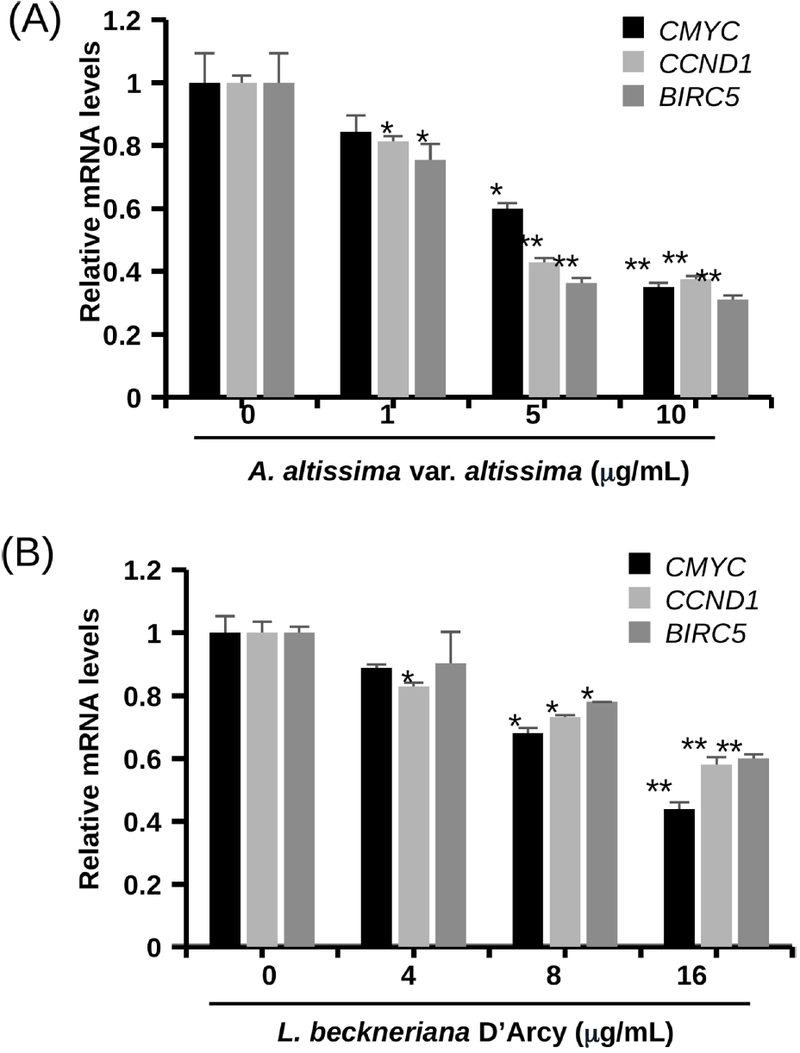
Effects of AAE and LBE on the mRNA expression of Wnt target genes in HCT116 cells. HCT116 cells were treated with the indicated concentrations of (A) AAE or (B) LBE for 12 h. The mRNA levels of CMYC, CCND1, and BIRC5 were determined by real-time PCR, normalized to those of ACTB, and are expressed relative to those of the vehicle-treated control. The data are expressed as the mean values ± SD (n = 3) and are representative of three separate determinations. *p < 0.05 and **p < 0.01 compared with the vehicle-treated control.
Consistent with these transcriptional changes, Western blot analysis after 24 h of treatment showed that AAE decreased the protein levels of c-Myc, cyclin D1, and survivin, with the most pronounced reduction observed for survivin at 5 and 10 μg/mL (Fig. 5A and C). LBE similarly reduced the levels of the three Wnt target proteins, at 16 μg/mL (Fig. 5B and D). Densitometric analysis of three independent experiments, in which the band intensities were normalized to those of β-actin, confirmed these reductions: at 10 μg/mL, AAE decreased the relative levels of c-Myc, cyclin D1, and survivin to 0.62, 0.39, and 0.22 of those of the control, respectively, and at 16 μg/mL, LBE decreased them to 0.15, 0.18, and 0.43, respectively (p < 0.01; Fig. 5C and D). Taken together, the reporter gene, real-time PCR, and Western blot data indicate that both extracts suppress Wnt/β-catenin signaling at the level of β-catenin/TCF-dependent transcription, and that this suppression is propagated to the expression of the downstream target proteins.
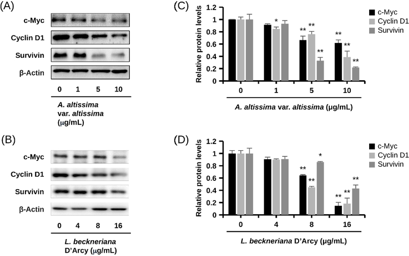
Effects of AAE and LBE on the expression of Wnt target proteins in HCT116 cells. HCT116 cells were treated with the indicated concentrations of (A) AAE or (B) LBE for 24 h, and the protein levels of c-Myc, cyclin D1, and survivin were determined by Western blot analysis. β-Actin was used as an internal standard. The blots are representative of three separate experiments. (C, D) The band intensities of c-Myc, cyclin D1, and survivin in (A) and (B), respectively, were quantified by densitometry, normalized to those of β-actin, and are expressed relative to those of the vehicle-treated control. The data are expressed as the mean values ± SD of three independent experiments. *p < 0.05 and **p < 0.01 compared with the vehicle-treated control.
The present findings are in line with previous reports in which natural products that suppress β-catenin/TCF transcription also reduced the expression of c-Myc, cyclin D1, and survivin and inhibited the proliferation of HCT116 cells.10–12 For A. altissima, previous phytochemical and pharmacological studies have identified quassinoids, in particular ailanthone, as the principal antitumor constituents,13,14 and ailanthone has been reported to inhibit the growth of human colorectal cancer cells through the suppression of STAT3 signaling.15 The strong inhibition of endogenous TOPflash activity by AAE observed in the present study indicates that this plant also affects the Wnt/β-catenin pathway, although the constituents responsible remain to be identified by bioassay-guided fractionation.
Because crude 95% ethanol extracts were used in the present study, the chemical composition of the tested materials should be considered when interpreting these results. Previous phytochemical studies of A. altissima have reported quassinoids, alkaloids, triterpenoids, phenylpropanoids, and flavonoids from various parts of the plant.13 In the leaves, which were included in the plant material used here, glycosides of the flavonols quercetin and kaempferol, with quercetin-3-O-glucoside as the predominant flavonoid, together with glycosides of the flavones apigenin and luteolin and phenolic acids such as caffeic and chlorogenic acids, have been identified by HPLC analysis.18 Among the known constituents, the quassinoid ailanthone is a plausible contributor to the potent activity of AAE, given its strong antiproliferative activity against various cancer cells, including colorectal cancer cells.14,15 Ailanthone has also recently been shown to suppress a HIF-1α/LINC01956/FUS axis that promotes the nuclear translocation of β-catenin in glioblastoma cells,19 indicating that it can modulate β-catenin signaling, albeit indirectly. It should be noted, however, that some quassinoids, such as brusatol, act as global inhibitors of protein synthesis and preferentially deplete short-lived proteins.20 Because c-Myc, cyclin D1, and survivin are short-lived proteins, such a mechanism might contribute to the reduction of their protein levels; nevertheless, the down-regulation of the corresponding transcripts after 12 h of treatment (Fig. 4) indicates that AAE acts, at least in part, at the transcriptional level. In addition, quercetin, the aglycone of the predominant leaf flavonoid, has been reported to inhibit β-catenin/TCF transcriptional activity in SW480 colorectal cancer cells by disrupting the binding of TCF complexes to their DNA-binding sites,21 and quercetin and its glycosides may therefore also contribute to the activity of AAE.
Lycianthes beckneriana, a taxonomically unrelated Solanaceae species, showed both potent growth inhibition and suppression of β-catenin/TCF transcription, and the concordant decreases in the CMYC, CCND1, and BIRC5 transcripts and in their encoded proteins support a mechanistic link between the two activities. For LBE, the IC50 value for the inhibition of TOPflash activity at 24 h (12.67 μg/mL) was higher than that for the inhibition of cell growth at 72 h (6.27 μg/mL). Since the two assays were performed after different exposure periods, these values are not directly comparable, and the data are most consistently interpreted as an early suppression of β-catenin-dependent transcription that translates into progressive growth inhibition during prolonged exposure. In contrast to A. altissima, no phytochemical or biological study of L. beckneriana has been reported to date. Preliminary phytochemical screening of another member of the genus, Lycianthes lycioides, has indicated the presence of alkaloids, phenolic compounds, flavonoids, saponins, triterpenes, and steroids,22 and these compound classes, which are also widely distributed in other Solanaceae plants, are candidate constituents of LBE. Because the extracts were available only in limited amounts (Table 1), the isolation and structural identification of their constituents could not be performed in the present study. Chemical profiling of AAE and LBE by LC-MS, together with bioassay-guided fractionation using the TOPflash reporter gene assay, will therefore be required to identify the compounds responsible for the observed activities.
Some limitations of the present study should be noted. Because both extracts showed considerable antiproliferative activity, a contribution of nonspecific cytotoxicity to the reduction of reporter activity must be considered. Several observations argue against cytotoxicity as the sole explanation: firefly luciferase activity was normalized to that of the cotransfected Renilla luciferase, which corrects for differences in cell number and transfection efficiency; the reporter gene assays were performed after 24 h of treatment, a much shorter exposure than that used in the 72-h growth assay; and the mRNA levels of the endogenous Wnt target genes were already decreased after 12 h of treatment. Nevertheless, because cell viability was not determined under the conditions of the reporter gene assays, a partial contribution of nonspecific cytotoxicity or of a general inhibition of transcription or translation to the effects observed at the highest concentrations cannot be completely excluded. The specificity of the inhibition should therefore be confirmed with the isolated active constituents, for example by determining cell viability in parallel and by comparison with β-catenin-independent reporter systems.
In conclusion, the 95% ethanol extracts of A. altissima var. altissima and L. beckneriana D’Arcy inhibited the growth of HCT116 human colorectal cancer cells and suppressed β-catenin/TCF-responsive transcription both in ectopically activated HEK293 cells and in HCT116 cells with constitutively active Wnt signaling. These effects were accompanied by the down-regulation of the Wnt target genes CMYC, CCND1, and BIRC5 and of the c-Myc, cyclin D1, and survivin proteins. AAE and LBE are therefore promising starting materials for the bioassay-guided identification of natural product inhibitors of Wnt/β-catenin signaling in colorectal cancer.
Acknowledgments
This research was supported by the National Research Foundation of Korea (NRF) grant funded by the Korea government (No. RS-2025-00518637) and was supported by Global – Learning & Academic research institution for Master’s·PhD students, and Postdocs (G-LAMP) Program of the NRF grant funded by the Ministry of Education (No. RS-2025-25442355).
Conflicts of Interest
The authors declare that they have no conflicts of interest.
References
-
Bray, F.; Laversanne, M.; Sung, H.; Ferlay, J.; Siegel, R. L.; Soerjomataram, I.; Jemal, A. CA Cancer J. Clin. 2024, 74, 229–263.
[https://doi.org/10.3322/caac.21834]

-
Dekker, E.; Tanis, P. J.; Vleugels, J. L. A.; Kasi, P. M.; Wallace, M. B. Lancet 2019, 394, 1467–1480.
[https://doi.org/10.1016/S0140-6736(19)32319-0]
-
Clevers, H. Cell 2006, 127, 469–480.
[https://doi.org/10.1016/j.cell.2006.10.018]
-
Clevers, H.; Nusse, R. Cell 2012, 149, 1192–1205.
[https://doi.org/10.1016/j.cell.2012.05.012]
-
MacDonald, B. T.; Tamai, K.; He, X. Dev. Cell 2009, 17, 9–26.
[https://doi.org/10.1016/j.devcel.2009.06.016]
-
Morin, P. J.; Sparks, A. B.; Korinek, V.; Barker, N.; Clevers, H.; Vogelstein, B.; Kinzler, K. W. Science 1997, 275, 1787–1790.
[https://doi.org/10.1126/science.275.5307.1787]
-
He, T. C.; Sparks, A. B.; Rago, C.; Hermeking, H.; Zawel, L.; da Costa, L. T.; Morin, P. J.; Vogelstein, B.; Kinzler, K. W. Science 1998, 281, 1509–1512.
[https://doi.org/10.1126/science.281.5382.1509]
-
Tetsu, O.; McCormick, F. Nature 1999, 398, 422–426.
[https://doi.org/10.1038/18884]
-
Kim, P. J.; Plescia, J.; Clevers, H.; Fearon, E. R.; Altieri, D. C. Lancet 2003, 362, 205–209.
[https://doi.org/10.1016/S0140-6736(03)13910-4]
-
Lee, M. A.; Park, H. J.; Chung, H. J.; Kim, W. K.; Lee, S. K. J. Nat. Prod. 2013, 76, 1278–1284.
[https://doi.org/10.1021/np400216m]
-
Kim, W. K.; Bach, D. H.; Ryu, H. W.; Oh, J.; Park, H. J.; Hong, J. Y.; Song, H. H.; Eum, S.; Bach, T. T.; Lee, S. K. Phytomedicine 2017, 34, 136–142.
[https://doi.org/10.1016/j.phymed.2017.08.008]
-
Kim, W. K.; Byun, W. S.; Chung, H. J.; Oh, J.; Park, H. J.; Choi, J. S.; Lee, S. K. Biochem. Pharmacol. 2018, 152, 71–83.
[https://doi.org/10.1016/j.bcp.2018.03.009]
-
Li, X.; Li, Y.; Ma, S.; Zhao, Q.; Wu, J.; Duan, L.; Xie, Y.; Wang, S. J. Ethnopharmacol. 2021, 275, 114121.
[https://doi.org/10.1016/j.jep.2021.114121]
-
Bailly, C. Phytother. Res. 2020, 34, 2203–2213.
[https://doi.org/10.1002/ptr.6681]
-
Ding, H.; Yu, X.; Yan, Z. Int. J. Mol. Med. 2022, 49, 21.
[https://doi.org/10.3892/ijmm.2021.5076]
-
Skehan, P.; Storeng, R.; Scudiero, D.; Monks, A.; McMahon, J.; Vistica, D.; Warren, J. T.; Bokesch, H.; Kenney, S.; Boyd, M. R. J. Natl. Cancer Inst. 1990, 82, 1107–1112.
[https://doi.org/10.1093/jnci/82.13.1107]
-
Livak, K. J.; Schmittgen, T. D. Methods 2001, 25, 402–408.
[https://doi.org/10.1006/meth.2001.1262]
-
Poljuha, D.; Sladonja, B.; Šola, I.; Dudaš, S.; Bilić, J.; Rusak, G.; Motlhatlego, K. E.; Eloff, J. N. Nat. Prod. Commun. 2017, 12, 1609–1612.
[https://doi.org/10.1177/1934578X1701201021]
-
Deng, X.; Zhang, Q.; Jin, F.; Lu, F.; Duan, G.; Han, L.; Zhu, M.; Yang, Z.; Zhang, G. Cancer Cell Int. 2024, 24, 397.
[https://doi.org/10.1186/s12935-024-03594-w]
-
Vartanian, S.; Ma, T. P.; Lee, J.; Haverty, P. M.; Kirkpatrick, D. S.; Yu, K.; Stokoe, D. Mol. Cell. Proteomics 2016, 15, 1220–1231.
[https://doi.org/10.1074/mcp.M115.055509]
-
Park, C. H.; Chang, J. Y.; Hahm, E. R.; Park, S.; Kim, H. K.; Yang, C. H. Biochem. Biophys. Res. Commun. 2005, 328, 227–234.
[https://doi.org/10.1016/j.bbrc.2004.12.151]
-
Soto Vásquez, M. R. Arnaldoa 2014, 21, 91–104.
[https://doi.org/10.3726/85617_91]