
A New Galloylated Megastigmane Glycoside from the Leaves of Psidium guajava and Antimicrobial Activity of the Isolated Compounds
Abstract
A new galloylated megastigmane glycoside, (3R,9R)-3-hydroxy-7,8-didehydro-β-ionol 9-O-(6′-O-galloyl)-β-D-glucopyranoside (1), together with four known compounds, mallophenol B (2), garcimangosone D (3), quercetin-3-O-α-L-arabinofuranoside (4), and quercetin-3-O-α-L-arabinopyranoside (5), was isolated from the leaves of Psidium guajava L. (Myrtaceae). Their structures were elucidated by NMR spectroscopy and comparison with published data, supported by HR-ESI-MS data where available. The isolated compounds were evaluated for antimicrobial activity against three Gram-positive bacteria, three Gram-negative bacteria, and Candida albicans. Compound 1 showed the strongest activity among the isolates, with MIC values of 64 μg/mL against Staphylococcus aureus and C. albicans. Compounds 2–5 were less active, and none of the compounds inhibited the tested Gram-negative bacteria at concentrations up to 256 μg/mL. These findings expand the chemical diversity of P. guajava leaves and provide compound-level information on their antimicrobial properties.
Keywords:
Psidium guajava, Myrtaceae, Megastigmane glycoside, Antimicrobial activityIntroduction
Antimicrobial resistance (AMR) is a major global health problem that reduces the effectiveness of anti-infective therapy and increases morbidity and mortality. In 2021, bacterial AMR was associated with approximately 4.71 million deaths worldwide, including 1.14 million deaths directly attributable to resistance.1 The World Health Organization’s 2025 global surveillance report further indicated that approximately one in six laboratory-confirmed bacterial infections caused by common pathogens in 2023 were resistant to antibiotic treatment.2 These trends support continued efforts to identify structurally diverse antimicrobial agents, including natural products from medicinal plants.
Psidium guajava L. (Myrtaceae), commonly known as guava, is cultivated in tropical and subtropical regions and has a long history of medicinal use.3 Preparations of the leaves have traditionally been used for diarrhea, dysentery, gastrointestinal disorders, cough, oral ulcers, and wound care.4 Antimicrobial effects have been reported for guava leaf extracts against several bacterial and fungal species,5,6 and activity has also been described for the leaf essential oil and its volatile constituents.7 These observations indicate that the leaves are a useful source for investigating individual metabolites that may contribute to the reported biological effects.
Phytochemical studies of P. guajava leaves have revealed phenolic compounds, flavonoids, triterpenoids, benzophenones, and meroterpenoids.8–12 Although the chemistry and antimicrobial properties of crude preparations have been investigated extensively, fewer studies have examined the activities of purified leaf constituents. In the present study, compounds were isolated from P. guajava leaves, characterized by spectroscopic methods, and evaluated for antimicrobial activity against selected bacteria and C. albicans.
Experimental
General experimental procedures – Preparative HPLC was carried out with an Agilent 1100 HPLC system (Agilent Technologies). UV spectra were recorded using the diode-array detector of the Agilent 1100 HPLC system (Agilent Technologies), with detection wavelengths set at 205, 230, 254, and 280 nm. Silica gel (Kieselgel 60, 230–400 mesh, Merck), Sephadex LH-20 (Merck), and YMC RP-18 resin (YMC Co. Ltd.) were used for column chromatography. NMR spectra were recorded on a Bruker AVANCE NEO 600 spectrometer at 600 MHz for 1H and 150 MHz for 13C. Optical rotations were measured using a Jasco P-2000 digital polarimeter. HR-ESI-MS spectra were acquired using a Thermo Scientific Orbitrap Exploris 240 mass spectrometer equipped with an electrospray ionization (ESI) source. GC analyses were performed using a Shimadzu GC-2010 gas chromatograph (Shimadzu Corporation).
Plant materials – Leaves of P. guajava were collected in Lao Cai, Vietnam, in May 2024. The plant was identified by Dr. Bui Van Thanh, Institute of Biology, VAST. Voucher material was deposited in the Institute of Biology, VAST, under the accession number PG2405.
Extraction and isolation – Dried leaves of P. guajava (6.0 kg) were sonicated twice with MeOH (15 L per extraction) for 2 h at 45°C. Evaporation of the extracts under reduced pressure yielded the MeOH extract (PGL, 1.1 kg). The residue was dispersed in water (1.5 L) and sequentially extracted with n-hexane and ethyl acetate. After solvent removal, the n-hexane and ethyl acetate layers gave PGL1 (75 g) and PGL2 (109 g), respectively; the residual water layer was retained as PGL3.
PGL3 was subjected to Diaion HP-20 column chromatography, beginning with water and followed by 25, 75, and 100% MeOH in water, to obtain PGL3A–PGL3C. PGL3A was separated on silica gel with CH2Cl2–MeOH (20:1, 10:1, 5:1, and 2.5:1, v/v) to yield PGL3A1–PGL3A4. PGL3A2 was further chromatographed on silica gel with CH2Cl2–acetone–H2O (1:1.5:0.1, v/v/v), followed by Sephadex LH-20 (MeOH–H2O, 9:1, v/v) and preparative HPLC using 25% ACN in water, to afford compound 4 (12.0 mg, tR = 32.3 min). PGL3B was divided into PGL3B1–PGL3B4 by silica gel chromatography. PGL3B2 was separated with CH2Cl2–acetone–H2O (1:1:0.05, v/v/v), and PGL3B2C was subsequently chromatographed on RP-18 with acetone–H2O (1:2, v/v). Sephadex LH-20 chromatography (MeOH–H2O, 9:1, v/v) followed by preparative HPLC using 20% ACN in water afforded compound 3 (4.2 mg, tR = 36.9 min). PGL3B3 was successively fractionated on silica gel [CH2Cl2–acetone–H2O (1:1.5:0.1, v/v/v)], RP-18 [acetone–H2O (1:2, v/v)], and Sephadex LH-20 [MeOH–H2O (9:1, v/v)] to give PGL3B3A–PGL3B3C. Preparative HPLC of PGL3B3A with 22.5% ACN in water yielded compounds 1 (4.2 mg, tR = 42.4 min) and 2 (6.0 mg, tR = 27.4 min), whereas PGL3B3C afforded compound 5 (3.8 mg, tR = 54.9 min) under the same HPLC conditions.
Acid hydrolysis – Compound 1 (3.0 mg) was hydrolyzed with 1.0 N HCl in dioxane–H2O (1:1, v/v) at 80°C for 3 h. After cooling to room temperature, the acidic solution was neutralized with Ag2CO3 and evaporated to dryness under a stream of N2. The residue was partitioned with H2O and CHCl3, and the aqueous fraction was collected and dried under N2. The resulting sugar residue was dissolved in anhydrous pyridine (0.1 mL), treated with L-cysteine methyl ester hydrochloride in pyridine (0.06 M, 0.1 mL), and maintained at 60°C for 2 h. Subsequently, trimethylsilylimidazole (0.1 mL) was added, and the reaction was continued at 60°C for 1.5 h to derivatize the sugars. After removing the solvent, the derivatized sugars were partitioned with n-hexane and H2O (0.1 mL each), and the n-hexane layer was subjected to GC analysis using a DB-5 column (0.32 mm × 30 m) equipped with a flame ionization detector (FID). The column, injector, and detector temperatures were maintained at 210, 270, and 300°C, respectively, with He as the carrier gas. The derivatives of authentic D-glucose and L-glucose exhibited retention times (tR) of 14.11 and 14.26 min, respectively. The derivatized sugar released from compound 1 showed a peak at tR = 14.12 min, consistent with authentic D-glucose (14.11 min) and distinct from L-glucose (14.26 min), supporting the D-glucose assignment.
(3R,9R)-3-hydroxy-7,8-didehydro-β-ionol 9-O-(6′-O-galloyl)-β-D-glucopyranoside (1) – White amorphous 25 powder; –8.0 (c 0.1, MeOH); C26H34O11, HR-ESI-MS m/z: 545.1995 [M + Na]+ (calcd for [C26H34O11Na]+, 545.1993); UV (MeOH) λmax 218 and 277 nm; 1H- and 13C-NMR (CD3OD): see Table 1.

1H- and 13C-NMR data for compound 1
Mallophenol B (2) – White amorphous powder; +14.0 (c 0.1, MeOH); C26H34O12, due to the limited amount of remaining sample, HR-ESI-MS data could not be obtained; 1H-NMR (CD3OD, 600 MHz) δH: 7.10 (2H, s, H-2′′/6′′), 5.83 (1H, d, J = 1.2 Hz, H-4), 5.82 (2H, overlapped, H-7/H-8), 4.48 (1H, dd, J = 2.4, 12.0 Hz, Hb-6′), 4.43 (1H, d, J = 7.8 Hz, H-1′), 4.40 (1H, m, H-9), 4.38 (1H, dd, J = 5.4, 12.0 Hz, Ha-6′), 3.52 (1H, m, H-5′), 3.46 (1H, t, J = 9.0 Hz, H-4′), 3.41 (1H, t, J = 9.0 Hz, H-3′), 3.24 (1H, dd, J = 7.8, 9.0 Hz, H-2′), 2.43 (1H, d, J = 16.8 Hz, Hb-2), 2.11 (1H, d, J = 16.8 Hz, Ha-2), 1.86 (3H, d, J = 1.2 Hz, H-11), 1.30 (3H, d, J = 6.6 Hz, H-10), 1.01 (3H, s, H-12), and 0.97 (3H, s, H-13). 13C-NMR (CD3OD, 150 MHz) δC: 201.3 (C-3), 168.4 (C-5, C-7′′), 146.5 (C-3′′/5′′), 139.9 (C-4′′), 134.9 (C-7), 131.6 (C-8), 127.2 (C-4), 121.5 (C-1′′), 110.3 (C-2′′/6′′), 103.1 (C-1′), 78.0 (C-3′), 77.1 (C-9), 75.5 (C-5′), 75.2 (C-2′), 71.5 (C-4′), 64.7 (C-6′), 50.7 (C-2), 42.5 (C-1), 24.6 (C-13), 23.4 (C-12), 21.2 (C-10), and 19.6 (C-11).
Garcimangosone D (3) – Yellow powder; –31.0 (c 0.1, MeOH); C19H20O9, HR-ESI-MS m/z: 415.0998 [M + Na]+ (calcd for [C19H20O9Na]+, 415.1000); 1H-NMR (CD3OD, 600 MHz) δH: 7.71 (2H, dd, J = 1.2, 8.4 Hz, H-2′/6′), 7.54 (1H, t, J = 7.2 Hz, H-4′), 7.44 (2H, t, J = 7.8 Hz, H-3′/5′), 6.23 (1H, d, J = 1.8 Hz, H-5), 6.09 (1H, d, J = 1.8 Hz, H-3), 4.84 (1H, m, H-1′′), 3.87 (1H, dd, J = 2.4, 12.0 Hz, Hb-6′′), 3.67 (1H, dd, J = 6.0, 12.0 Hz, Ha-6′′), 3.35 (1H, m, H-3′′), 3.32 (1H, m, H-5′′), 3.24 (1H, t, J = 9.0 Hz, H-4′′), and 2.85 (1H, dd, J = 7.2, 9.0 Hz, H-2′′). 13C-NMR (CD3OD, 150 MHz) δC: 199.5 (C-7), 164.1 (C-4), 162.1 (C-2), 159.7 (C-6), 141.7 (C-1′), 133.1 (C-4′), 130.1 (C-2′/6′), 129.0 (C-3′/5′), 109.1 (C-1), 101.7 (C-1′′), 98.2 (C-3), 95.8 (C-5), 78.2 (C-5′′), 77.8 (C-3′′), 74.6 (C-2′′), 71.1 (C-4′′), and 62.5 (C-6′′).
Quercetin-3-O-α-L-arabinofuranoside (4) – Yellow 25 powder; –17.0 (c 0.1, MeOH); C20H18O11, HR-ESI-MS m/z: 457.0742 [M + Na]+ (calcd for [C20H18O11Na]+, 457.0741); 1H-NMR (CD3OD, 600 MHz) δH: 7.54 (1H, d, J = 1.8 Hz, H-2′), 7.51 (1H, dd, J = 1.8, 8.4 Hz, H-6′), 6.91 (1H, d, J = 8.4 Hz, H-5′), 6.40 (1H, d, J = 1.8 Hz, H-8), 6.22 (1H, d, J = 1.8 Hz, H-6), 5.49 (1H, s, H-1′′), 4.35 (1H, d, J = 1.8 Hz, H-2′′), 3.93 (1H, dd, J = 2.4, 5.4 Hz, H-3′′), 3.89 (1H, q, J = 4.2 Hz, H-4′′), 3.54 (1H, dd, J = 4.2, 12.0 Hz, Hb-5′′), and 3.52 (1H, dd, J = 4.8, 12.0 Hz, Ha-5′′). 13C-NMR (CD3OD, 150 MHz) δC: 180.0 (C-4), 166.1 (C-7), 163.0 (C-5), 159.3 (C-2), 158.6 (C-9), 149.8 (C-4′), 146.3 (C-3′), 134.9 (C-3), 123.1 (C-6′), 123.0 (C-1′), 116.9 (C-2′), 116.4 (C-5′), 109.5 (C-1′′), 105.6 (C-10), 99.9 (C-6), 94.8 (C-8), 88.0 (C-4′′), 83.3 (C-2′′), 78.7 (C-3′′), and 62.5 (C-5′′).
Quercetin-3-O-α-L-arabinopyranoside (5) – Yellow 25 powder; –23.0 (c 0.1, MeOH); C20H18O11, HR-ESI-MS m/z: 457.0743 [M + Na]+ (calcd for [C20H18O11Na]+, 457.0741); 1H-NMR (CD3OD, 600 MHz) δH: 7.76 (1H, d, J = 1.8 Hz, H-2′), 7.59 (1H, dd, J = 1.8, 8.4 Hz, H-6′), 6.89 (1H, d, J = 8.4 Hz, H-5′), 6.41 (1H, s, H-8), 6.22 (1H, d, J = 1.8 Hz, H-6), 5.17 (1H, d, J = 6.6 Hz, H-1′′), 3.92 (1H, dd, J = 6.6, 8.4 Hz, H-2′′), 3.83 (2H, m, H-4′′/Hb-5′′), 3.66 (1H, m, H-3′′), and 3.45 (1H, m, Ha-5′′). 13C-NMR (CD3OD, 150 MHz) δC: 178.1 (C-4), 164.7 (C-7), 161.6 (C-5), 157.3 (C-9), 157.0 (C-2), 148.6 (C-4′), 144.6 (C-3′), 134.3 (C-3), 121.7 (C-1′), 121.5 (C-6′), 116.1 (C-2′), 114.8 (C-5′), 104.2 (C-10), 103.3 (C-1′′), 98.5 (C-6), 93.4 (C-8), 72.7 (C-3′′), 70.7 (C-2′′), 67.7 (C-4′′), and 65.5 (C-5′′).
Antimicrobial assay – Compounds 1–5 were subjected to antimicrobial testing against three Gram-positive bacteria (Enterococcus faecalis ATCC 29212, Staphylococcus aureus ATCC 25923, and Bacillus cereus ATCC 14579), three Gram-negative bacteria (Escherichia coli ATCC 25922, Pseudomonas aeruginosa ATCC 27853, and Salmonella enterica ATCC 13076), and one yeast (Candida albicans ATCC 10231). The assay was performed in 96-well plates using a broth-dilution format based on a reported method with minor modifications.13 For each test well, 10 μL of compound solution in DMSO and 190 μL of a microorganism suspension (2 × 10⁵ CFU/mL) were combined. Plates were maintained at 37°C for 24 h, after which 10 μL of resazurin solution (0.01%) was added. The plates were further incubated for 1 h until a color change was observed, and the absorbance was subsequently measured at 600 nm. MIC was assigned to the lowest concentration producing at least 97% growth inhibition relative to the untreated control. Streptomycin served as the bacterial positive control, whereas cycloheximide was used for C. albicans.
Results and Discussion
Chromatographic separation of the aqueous fraction of P. guajava leaves afforded one new megastigmane glycoside (1) and four known compounds (2–5) (Fig. 1).
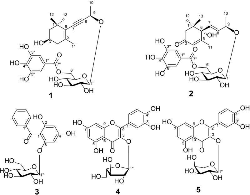
Chemical structures of compounds 1–5 isolated from P. guajava leaves.
Compound 1 was isolated as a white amorphous powder. The molecular formula was established as C26H34O11 from the sodium-adduct ion at m/z 545.1995 [M + Na]+ (calcd for [C26H34O11Na]+, 545.1993) in the HR-ESI-MS spectrum. The 1H-NMR spectrum exhibited a two-proton singlet at δH 7.10 (2H, s), assigned to the equivalent galloyl protons H-2′′ and H-6′′, together with an anomeric proton at δH 4.60 (1H, d, J = 7.8 Hz, H-1′), and four methyl signals at δH 1.05 (s, H-12), 1.11 (s, H-13), 1.51 (d, J = 6.6 Hz, H-10), and 1.81 (s, H-11). The 13C-NMR and HSQC data accounted for 26 carbons, including a galloyl carbonyl, six aromatic carbons, six glucosyl carbons, and 13 carbons attributable to a megastigmane unit. Comparison of the spectral features with those of the reported (3R,9R)-3-hydroxy-7,8-didehydro-β-ionyl 9-O-α-D-arabinopyranosyl-(1→6)-β-D-glucopyranoside14 showed a key difference at C-6′, where compound 1 bears a galloyl group. An HMBC cross-peak from H-6′ (δH 4.42 and 4.50) to the galloyl carbonyl C-7′′ (δC 168.4) assigned the galloyl substitution at C-6′ (Fig. 2). A further correlation from H-1′ (δH 4.60) to C-9 (δC 67.9) confirmed the 9-O-glucosyl linkage. Additional HMBC cross-peaks from H-2/H-4 to C-3, H-10 to C-8/C-9, H-11 to C-5/C-6, and H-12/H-13 to C-6 were consistent with the substitution pattern of the aglycone (Fig. 2). The large vicinal coupling between H-3 and H-4 (J = 9.6 Hz) supported an axial orientation of H-3; comparison with the related glucoside and synthetic stereoisomers supported the 3R,9R assignment.14,15 Accordingly, compound 1 was assigned as (3R,9R)-3-hydroxy-7,8-didehydro-β-ionol 9-O-(6′-O-galloyl)-β-D-glucopyranoside.
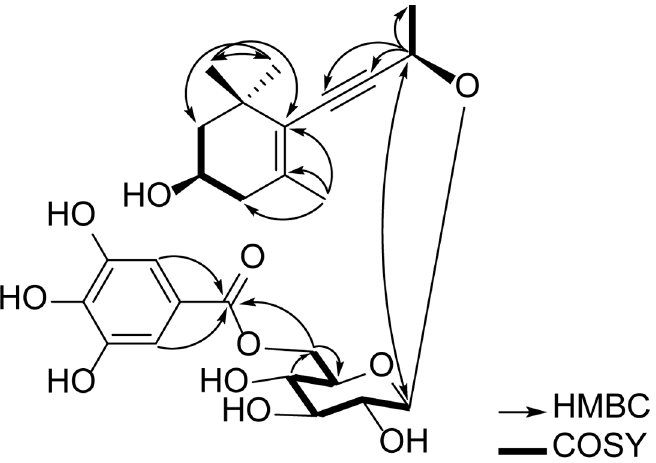
Key HMBC and COSY correlations of compound 1.
Compound 2 was characterized by a galloyl singlet at δH 7.10 (2H) and a β-glucosyl anomeric doublet at δH 4.43 (1H, J = 7.8 Hz), along with signals attributable to a blumenol A-type aglycone.16 The glucose C-6′ signal appeared at δC 64.7, downfield from that expected for an unsubstituted glucosyl hydroxymethyl carbon, supporting galloylation at C-6′. These spectroscopic features supported its assignment as mallophenol B.17
Compound 3 showed signals characteristic of a benzophenone framework together with a glucopyranosyl moiety. The carbon data comprised one ketone carbonyl, twelve aromatic carbons corresponding to two benzene rings, and six resonances from a hexose. Agreement with published reference data supported assignment of 3 as garcimangosone D.18
Compounds 4 and 5 shared the characteristic aromatic resonances of quercetin glycosides, whereas their sugar signals differentiated an α-L-arabinofuranosyl unit in 4 from an α-L-arabinopyranosyl unit in 5. These data supported assignment of the two compounds as quercetin-3-O-α-L-arabinofuranoside19 and quercetin-3-O-α-L-arabinopyranoside,20 respectively.
Antimicrobial activities of the isolated compounds are summarized in Table 2. Compound 1 showed the strongest activity, inhibiting S. aureus and C. albicans at 64 μg/mL and E. faecalis and B. cereus at 128 μg/mL. Compounds 2 and 3 gave MIC values of 128–256 μg/mL against the susceptible Gram-positive strains and 128 μg/mL against C. albicans. Compounds 4 and 5 showed weaker activity against the Gram-positive bacteria (MIC = 256 μg/mL) but inhibited C. albicans at 128 μg/mL. No activity was detected against E. coli, P. aeruginosa, or S. enterica at the highest concentration examined (256 μg/mL). Overall, the activity profile was selective and modest, and compound 1 was the most active of the tested isolates.
Antimicrobial activities of compounds 1–5
Garcimangosone D (3) and quercetin-3-O-α-L-arabinopyranoside (5) were previously reported to inhibit E. coli and S. aureus, with stronger activity against S. aureus.21 Quercetin-3-O-α-L-arabinofuranoside (4) has also shown antifungal activity against Candida spp., with reported MIC values of 2–32 μg/mL in a study of Myrcia tomentosa leaves.22 To the best of our knowledge, no previous antibacterial or antifungal activity of mallophenol B (2) has been reported.
Megastigmane glycosides have also been evaluated in antimicrobial studies involving E. faecalis, S. aureus, and C. albicans, although activity varies with structure.23 A 6′-O-galloylated megastigmane glucoside from Gymnosporia senegalensis has likewise been evaluated for antibacterial and antifungal activity,24 and antimicrobial effects of megastigmane glycosides have been summarized in a recent review.25 In the present series, both compounds 1 and 2 are 6′-O-galloylated megastigmane glucosides; however, compound 1 showed twofold lower MIC values than compound 2 against both S. aureus and C. albicans (64 μg/mL for compound 1 vs 128 μg/mL for compound 2). This finding suggests that differences in the megastigmane aglycone, rather than galloylation alone, may contribute to antimicrobial activity. However, given the limited number of compounds examined, this structure–activity relationship should be considered preliminary.
Overall, investigation of P. guajava leaves yielded a new galloylated megastigmane glycoside (1) along with four previously reported compounds (2–5). Antimicrobial testing showed preferential activity toward Gram-positive bacteria and C. albicans, while compound 1 displayed the lowest MIC values in the series. These results add a galloylated megastigmane glycoside to the reported chemistry of P. guajava and define the antimicrobial profile of the isolated leaf constituents.
Acknowledgments
This research was funded by the Vietnam Academy of Science and Technology (Grant No. UQĐTCB.06/24-26).
Conflicts of Interest
The authors declare that they have no conflicts of interest.
References
- GBD 2021 Antimicrobial Resistance Collaborators. Lancet 2024, 404, 1199–1226.
- World Health Organization. Global antibiotic resistance surveillance report 2025; World Health Organization: Geneva, 2025.
- Gutiérrez, R. M. P.; Mitchell, S.; Solis, R. V. J. Ethnopharmacol. 2008, 117, 1–27.
-
Díaz-de-Cerio, E.; Verardo, V.; Gómez-Caravaca, A. M.; Fernández-Gutiérrez, A.; Segura-Carretero, A. Int. J. Mol. Sci. 2017, 18, 897.
[https://doi.org/10.3390/ijms18040897]

- Das, M.; Goswami, S. Int. J. Health Sci. Res. 2019, 9, 39–45.
-
Dhiman, A.; Nanda, A.; Ahmad, S.; Narasimhan, B. J. Pharm. Bioallied Sci. 2011, 3, 226–229.
[https://doi.org/10.4103/0975-7406.80776]
-
Soliman, F. M.; Fathy, M. M.; Salama, M. M.; Saber, F. R. Bull. Fac. Pharm. Cairo Univ. 2016, 54, 219–225.
[https://doi.org/10.1016/j.bfopcu.2016.06.003]
-
Begum, S.; Hassan, S. I.; Ali, S. N.; Siddiqui, B. S. Nat. Prod. Res. 2004, 18, 135–140.
[https://doi.org/10.1080/14786410310001608019]
-
Díaz-de-Cerio, E.; Gómez-Caravaca, A. M.; Verardo, V.; Fernández-Gutiérrez, A.; Segura-Carretero, A. J. Funct. Foods 2016, 22, 376–388.
[https://doi.org/10.1016/j.jff.2016.01.040]
-
Matsuzaki, K.; Ishii, R.; Kobiyama, K.; Kitanaka, S. J. Nat. Med. 2010, 64, 252–256.
[https://doi.org/10.1007/s11418-010-0400-2]
-
Shao, M.; Wang, Y.; Huang, X.-J.; Fan, C.-L.; Zhang, Q.-W.; Zhang, X.-Q.; Ye, W.-C. J. Asian Nat. Prod. Res. 2012, 14, 348–354.
[https://doi.org/10.1080/10286020.2011.653964]
-
Shao, M.; Wang, Y.; Liu, Z.; Zhang, D.-M.; Cao, H.-H.; Jiang, R.- W.; Fan, C.-L.; Zhang, X.-Q.; Chen, H.-R.; Yao, X.-S. Org. Lett. 2010, 12, 5040–5043.
[https://doi.org/10.1021/ol102179u]
-
Gharat, Y. P.; Abdelhamid, A. G.; Yousef, A. E. Front. Cell. Infect. Microbiol. 2025, 15, 1662291.
[https://doi.org/10.3389/fcimb.2025.1662291]
-
Shao, J.-H.; Chen, J.; Zhao, C.-C.; Shen, J.; Liu, W.-Y.; Gu, W.-Y.; Li, K.-H. Nat. Prod. Res. 2019, 33, 2662–2667.
[https://doi.org/10.1080/14786419.2018.1466130]
-
Yamano, Y.; Watanabe, Y.; Watanabe, N.; Ito, M. J. Chem. Soc., Perkin Trans. 1 2002, 2833–2844.
[https://doi.org/10.1039/b208758h]
- Erosa-Rejón, G.; Peña-Rodríguez, L. M.; Sterner, O. J. Mex. Chem. Soc. 2009, 53, 44–47.
-
Wei, K.; Li, W.; Koike, K.; Liu, L.; Fu, X.; Lin, L.; Chen, Y.; Nikaido, T. Chem. Pharm. Bull. 2004, 52, 776–779.
[https://doi.org/10.1248/cpb.52.776]
-
Huang, Y.-L.; Chen, C.-C.; Chen, Y.-J.; Huang, R.-L.; Shieh, B.-J. J. Nat. Prod. 2001, 64, 903–906.
[https://doi.org/10.1021/np000583q]
-
Rodrigues, E. D.; da Silva, D. B.; de Oliveira, D. C. R.; da Silva, G. V. J. Magn. Reson. Chem. 2009, 47, 1095–1100.
[https://doi.org/10.1002/mrc.2516]
-
Schieber, A.; Hilt, P.; Conrad, J.; Beifuss, U.; Carle, R. J. Sep. Sci. 2002, 25, 361–364.
[https://doi.org/10.1002/1615-9314(20020401)25:5/6<361::AID-JSSC361>3.0.CO;2-D]
-
Ukwueze, S. E.; Osadebe, P. O.; Okoye, F. B. Nat. Prod. Res. 2015, 29, 1728–1734.
[https://doi.org/10.1080/14786419.2014.1003188]
-
da Silva Sa, F. A.; De Paula, J. A. M.; Dos Santos, P. A.; de Almeida Ribeiro Oliveira, L.; de Almeida Ribeiro Oliveira, G.; Liao, L. M.; De Paula, J. R.; do Rosario Rodrigues Silva, M. Molecules 2017, 22, 1100.
[https://doi.org/10.3390/molecules22071100]
-
Nguyen, T. N.; Tam, L. T.; Pham Thi Mai, H.; Tran Thi Hong, H.; Ninh, T. N.; Cuong, D. V.; Nguyen Xuan, C.; Tran, H. Q. Nat. Prod. Res. 2023, 37, 2862–2870.
[https://doi.org/10.1080/14786419.2022.2138871]
-
Tatsimo Ndendoung, S. J.; Tamokou, J.-D.-D.; Toume, K.; Havyarimana, L.; Ekom, S. E.; Komatsu, K. J. Chem. Res. 2022, 46, 17475198211072498.
[https://doi.org/10.1177/17475198211072498]
-
Samra, R. M.; Othman, A.; Elsbaey, M.; Amen, Y.; Shimizu, K. Phytochem. Lett. 2024, 60, 19–89.
[https://doi.org/10.1016/j.phytol.2024.01.008]