
Demethoxycurcumin Suppresses Melanogenesis and Modulates the SOX10/CREB-MITF Axis in Melan-a Cells
Abstract
Natural products from plants, fungi, bacteria, and other organisms remain important sources of pharmacologically active compounds. Demethoxycurcumin (DC), a naturally occurring curcuminoid found in Curcuma aromatica (Zingiberaceae), was investigated for its anti-melanogenic activity and associated molecular changes in mouse melan-a cells. DC reduced cellular melanin content in a concentration-dependent manner, with an IC50 value of 17.1 μM, without reducing cell viability after 24 h at concentrations up to 30 μM. DC decreased the mRNA and protein expression of tyrosinase (TYR), tyrosinase-related protein-1 (TRP-1), and TRP-2, together with reduced microphthalmia-associated transcription factor (MITF) mRNA expression. Western blot analysis indicated reduced SOX10 protein abundance at the highest concentration examined and suggested increased ERK and AKT phosphorylation following DC treatment. In a reconstructed human melanized skin model, DC reduced melanin content by up to 24.4%. Collectively, these findings identify DC as an anti-melanogenic natural product whose activity is associated with modulation of the SOX10/CREB-MITF regulatory axis and reduced expression of melanogenic enzymes.
Keywords:
Demethoxycurcumin, MITF, Melan-a cells, MelanogenesisIntroduction
Melanin is synthesized by melanocytes and is a major determinant of skin pigmentation. The melanogenic pathway is primarily regulated by tyrosinase (TYR), which catalyzes the rate-limiting reactions of melanin biosynthesis, together with tyrosinase-related protein-1 (TRP-1) and TRP-2, which participate in downstream eumelanin synthesis.1–3
Microphthalmia-associated transcription factor (MITF) is a central transcriptional regulator of melanocyte differentiation and melanogenesis and regulates the expression of TYR, TRP-1, and TRP-2. MITF transcription is controlled by several lineage- and signal-dependent factors, including SRY-related HMG-box 10 (SOX10), paired box 3 (PAX3), and cAMP response element-binding protein (CREB). SOX10 and PAX3 regulate MITF transcription, whereas phosphorylated CREB promotes MITF transcription through binding to cAMP-responsive elements.4–6
MITF expression and activity are additionally influenced by intracellular signaling pathways, including ERK/MAPK and PI3K/AKT signaling. These pathways can regulate melanogenesis through modulation of MITF and its downstream melanogenic enzymes.2,7–9 Thus, evaluation of MITF transcription, its upstream transcriptional regulators, and associated intracellular signaling can help define the molecular actions of anti-melanogenic compounds.
Natural products from diverse biological sources, including microbial metabolites and plant-derived phytochemicals, have been extensively investigated as anti-melanogenic agents through direct modulation of tyrosinase activity and regulation of MITF-associated signaling pathways.10–12 Curcuminoids are diarylheptanoid constituents of Curcuma species and have attracted considerable interest because of their diverse biological activities.13
Curcumin has been reported to suppress melanogenesis in both B16F10 melanoma cells and cultured human melanocytes, accompanied by activation of ERK and AKT signaling and downregulation of MITF and melanogenic enzymes.14,15 Comparative studies of curcumin and structurally related curcuminoids have further demonstrated compound-dependent differences in anti-melanogenic activity.16 A curcumin derivative, J147, also suppressed melanogenesis through ERK-mediated regulation of MITF.17 Related anti-melanogenic effects have also been demonstrated in reconstructed three-dimensional human skin models.18 However, the molecular effects of demethoxycurcumin (DC) on melanogenesis have been less extensively characterized than those of curcumin. In particular, whether DC treatment is associated with modulation of upstream MITF regulators such as SOX10 and CREB remains unclear. Therefore, the present study investigated the anti-melanogenic activity of DC in melan-a cells, with particular emphasis on the SOX10/ CREB-MITF regulatory axis and associated ERK/AKT signaling. The anti-melanogenic activity of DC was further evaluated in a reconstructed human melanized skin model.
Experimental
Reagents and antibodies – RPMI 1640 medium, fetal bovine serum (FBS), antibiotic-antimycotic solution, and TRI reagent were obtained from Invitrogen (Grand Island, NY, USA). Bovine serum albumin (BSA), 3-(4,5-dimethylthiazol-2-yl)-2,5-diphenyltetrazolium bromide (MTT), bicinchoninic acid (BCA) reagent, dimethyl sulfoxide (DMSO), and other general reagents were purchased from Sigma-Aldrich (St. Louis, MO, USA).
Primary antibodies against tyrosinase (sc-7833), TRP-1 (sc-10443), TRP-2 (sc-10451), SOX10 (sc-17342), PAX3 (sc-7748), ERK (sc-94), phospho-ERK (sc-7383), and β-actin (sc-47778) were obtained from Santa Cruz Biotechnology (Santa Cruz, CA, USA) and used at 1:500 dilution. Primary antibodies against AKT (#9272) and phospho-AKT (Ser473; #9271) were obtained from Cell Signaling Technology (Beverly, MA, USA) and used at 1:1,000 dilution. The TransAM CREB DNA-binding assay kit was obtained from Active Motif Japan (Tokyo, Japan). Neoderm®-ME and Neoderm®-ED reconstructed human skin models were purchased from Tego Science (Seoul, Republic of Korea). Neoderm®-ME, which contains melanocytes, was used to evaluate the anti-melanogenic activity of DC in a reconstructed human skin model. Neoderm®-ED was used separately for tissue viability assessment as part of the original experimental design, whereas Neoderm®-ME was specifically used to evaluate melanin production in melanocyte-containing reconstructed skin.
Compound – Demethoxycurcumin (DC; Fig. 1A) was purchased from Sigma-Aldrich and dissolved in 100% DMSO to prepare a stock solution.
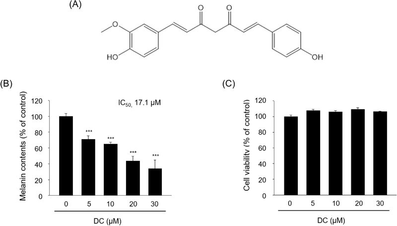
Inhibitory effect of demethoxycurcumin on melanin synthesis in melan-a cells. (A) Chemical structure of DC. (B) Cells were treated with 0, 5, 10, 20, or 30 µM DC for 48 h, and intracellular melanin content was measured. (C) Cell viability was assessed by MTT after 24 h of DC treatment. Data are presented as mean ± SD from three independent experiments (n = 3). For panel B, statistical significance was determined by one-way ANOVA followed by Dunnett’s multiple-comparison test versus the untreated control. ***p < 0.001 versus the untreated control.
Cell culture – Melan-a cells, originally established by Bennett et al.,19 were kindly provided by the Skin Research Institute, Amorepacific Corporation (Yongin, Republic of Korea). Cells were maintained in RPMI 1640 supplemented with 10% FBS, penicillin (100 units/mL), streptomycin (100 μg/mL), amphotericin B (250 ng/mL), and 200 nM 12-O-tetradecanoylphorbol-13-acetate (TPA) at 37°C in a humidified atmosphere containing 10% CO2.
Melanin content assay – Melan-a cells were seeded in 6-well plates at a density of 4 × 10⁵ cells/well. Forty-eight hours after seeding, the medium was replaced with medium containing the indicated concentrations of DC. After a further 48 h, cells were washed with Ca2+- and Mg2+-free phosphate-buffered saline (PBS), harvested, and pelleted. The pellets were solubilized in 0.1 mL of 1 N NaOH at 60°C for 10 min, and absorbance was measured at 475 nm. Melanin content was expressed as a percentage of the untreated control. For the reconstructed human skin assay, Neoderm®-ME tissues were maintained according to the manufacturer’s instructions, irradiated with 50 mJ/cm2 UVB every 3 days for a total of two exposures, and treated with DC for 4 days. Tissues were dissolved in 1 N NaOH, insoluble debris was removed by centrifugation at 13,000 rpm for 15 min, and absorbance of the supernatant was measured at 475 nm.
Cell viability assay – Cell viability was assessed by the MTT assay. MTT was added to a final concentration of 500 μg/mL, and samples were incubated for 3 h at 37°C. The resulting formazan was dissolved in DMSO, and absorbance was measured at 570 nm. Viability was expressed as a percentage of the untreated control. For a separate assessment of tissue viability, Neoderm®-ED tissues were treated with the indicated concentrations of DC for 3 days and analyzed by the MTT assay.
Western blot analysis – Melan-a cells were treated with the indicated concentrations of DC for the stated times, washed, and lysed. Protein concentrations were determined by the BCA assay. Equal amounts of protein were separated by 8–13% SDS-polyacrylamide gel electrophoresis and transferred to polyvinylidene difluoride membranes. Membranes were blocked with 5% BSA in Tris-buffered saline containing Tween 20, incubated with primary antibodies overnight at 4°C, and subsequently incubated with the appropriate horseradish peroxidase-conjugated secondary antibodies for 2 h at room temperature. Immunoreactive bands were detected using enhanced chemiluminescence and imaged with a LAS 4000 system (GE Healthcare, Little Chalfont, UK).
RNA extraction and quantitative real-time PCR – Total RNA was extracted using TRI reagent and reverse-transcribed using the Reverse Transcription System (Promega, Madison, WI, USA). Quantitative real-time PCR was performed with iQ™ SYBR® Green Supermix (Bio-Rad, Hercules, CA, USA). The thermal program consisted of 20 s at 95°C, followed by 40 cycles of 20 s at 95°C, 20 s at 56°C, and 30 s at 72°C, followed by 1 min at 95°C and 1 min at 55°C. Relative expression was calculated using the comparative Ct method with β-actin as the reference gene. Primer sequences are listed in Table 1.

Primer sequences used for quantitative real-time PCR
Nuclear extraction and phospho-CREB DNA-binding assay – Cells were washed with PBS, pelleted, and resuspended in lysis buffer containing 10 mM Tris-HCl (pH 8.0), 60 mM KCl, 1 mM EDTA, 1 mM dithiothreitol, 100 μM phenylmethylsulfonyl fluoride, and 1% NP-40. After 5 min on ice, lysates were centrifuged at 2,500 rpm for 4 min at 4°C. Nuclear pellets were washed with lysis buffer lacking NP-40 and resuspended in nuclear 420 mM NaCl, 1.5 mM MgCl2, 0.2 mM EDTA, and 25% glycerol. After incubation for 10 min at 4°C, samples were vortexed and centrifuged at 14,000 rpm for 5 min. Nuclear proteins (2 μg) were applied to wells coated with a double-stranded oligonucleotide containing the consensus CRE sequence (TGACGTCA). Bound phospho-CREB (Ser133) was detected using the TransAM assay according to the manufacturer’s instructions, and absorbance was measured at 450 nm.
Statistical analysis – Data are presented as mean ± standard deviation (SD) from three independent experiments unless otherwise indicated. For experiments involving multiple DC concentrations compared with a single untreated control, statistical significance was assessed by one-way analysis of variance (ANOVA) followed by Dunnett’s multiple-comparison test. For the reconstructed skin experiments in Fig. 5, comparisons between each DC-treated group and the untreated control were performed using Student’s t-test. Differences were considered statistically significant at p < 0.05.
Results and Discussion
DC decreased melanin content in melan-a cells in a concentration-dependent manner. At 5, 10, 20, and 30 μM, melanin content was decreased by 29.0, 35.3, 56.7, and 66.1%, respectively, compared with the untreated control, yielding an IC50 value of 17.1 μM (Fig. 1B). In contrast, DC did not reduce melan-a cell viability after 24 h of treatment at concentrations up to 30 μM (Fig. 1C).
To determine whether the reduction in melanin was associated with altered expression of melanogenic enzymes, TYR, TRP-1, and TRP-2 expressions were examined at the protein and mRNA levels. Western blot analysis showed an apparent reduction in TYR, TRP-1, and TRP-2 protein levels following DC treatment at both 24 and 48 h (Fig. 2A). At 24 h, TRP-1 mRNA was decreased by 14.7, 52.8, and 83.9%; TRP-2 mRNA was decreased by 13.9, 70.4, and 94.2%; and TYR mRNA was decreased by 4.5, 40.2, and 89.3% at 10, 20, and 30 μM DC, respectively (Fig. 2B). These findings indicate that the anti-melanogenic activity of DC is accompanied by reduced expression of melanogenic enzymes. MITF is a key transcriptional regulator of TYR, TRP-1, and TRP-2.1,2 MITF mRNA showed modest decreases at 10 and 20 μM DC and was markedly reduced by 83.1% at 30 μM DC after 24 h (Fig. 3A). Western blot analysis showed no apparent concentration-dependent change in PAX3, whereas SOX10 protein abundance appeared reduced at 30 μM DC (Fig. 3B). These findings are consistent with an association between SOX10 downregulation and reduced MITF transcription, although direct causality was not tested.
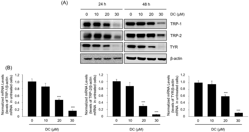
Effects of demethoxycurcumin on the expression of melanogenic enzymes in melan-a cells. (A) Cells were treated with 0, 10, 20, or 30 µM DC for 24 or 48 h, and TYR, TRP-1, and TRP-2 protein levels were examined by Western blotting. β-Actin was used as a loading control. (B) Cells were treated with DC for 24 h, and mRNA levels of TRP-1, TRP-2, and TYR were measured by quantitative real-time PCR and normalized to β-actin. Data are presented as mean ± SD from three independent experiments (n = 3). For panel B, statistical significance was determined by one-way ANOVA followed by Dunnett’s multiple-comparison test versus the untreated control. ***p < 0.001 versus the untreated control.
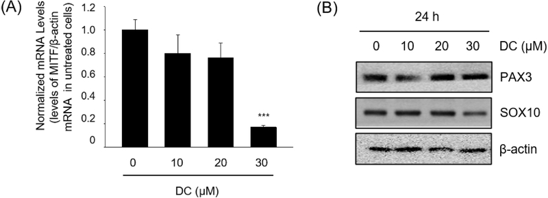
Effects of demethoxycurcumin on MITF mRNA expression and upstream transcriptional regulators. (A) MITF mRNA expression after treatment with 0–30 µM DC for 24 h, normalized to β-actin. Data are presented as mean ± SD from three independent experiments (n = 3). Statistical significance was determined by one-way ANOVA followed by Dunnett’s multiple-comparison test versus the untreated control. ***p < 0.001 versus the untreated control. (B) PAX3 and SOX10 protein expression after treatment with 0–30 µM DC for 24 h. β-Actin was used as a loading control.
Phospho-CREB DNA-binding activity was decreased by 22.7, 34.8, and 47.0% at 10, 20, and 30 μM DC, respectively (Fig. 4A). Because CREB contributes to MITF promoter activation,6,20 and suppression of cAMP/ PKA/CREB signaling has been associated with reduced melanogenesis by natural compounds,21 the reduction in phospho-CREB DNA-binding activity is consistent with the observed decrease in MITF mRNA expression, although a direct causal relationship was not tested.
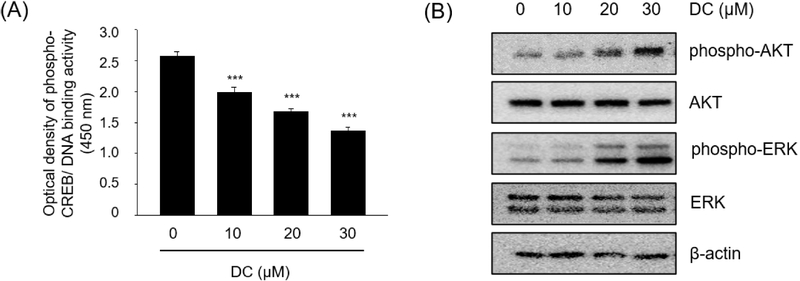
Effects of demethoxycurcumin on phospho-CREB DNA-binding activity and ERK/AKT signaling in melan-a cells. (A) Phospho-CREB DNA-binding activity was measured using an ELISA-based TransAM assay after treatment with 0–30 µM DC for 24 h. Data are presented as mean ± SD from three independent experiments (n = 3). Statistical significance was determined by one-way ANOVA followed by Dunnett’s multiple-comparison test versus the untreated control. ***p < 0.001 versus the untreated control. (B) Phospho-AKT, total AKT, phospho-ERK, and total ERK were examined after 24 h of DC treatment. β-Actin was used as a loading control.
Western blot analysis suggested increased phosphorylation of AKT and ERK following DC treatment, while total AKT and ERK levels appeared comparatively stable (Fig. 4B). This pattern is consistent with previous reports in which curcumin-associated suppression of melanogenesis was accompanied by ERK and AKT activation.14,15 Activation of ERK and/or AKT signaling has also been associated with reduced melanogenesis in response to other natural products.22,23 Thus, the changes in ERK and AKT phosphorylation associated with DC appear to overlap with those previously reported for curcumin. The present study further examined upstream regulators of MITF and showed that DC treatment was associated with reduced SOX10 abundance and phospho-CREB DNA-binding activity. However, because direct comparisons with curcumin or other curcuminoids and pathway inhibition, knockdown, or rescue experiments were not performed, the present data do not establish differences in relative potency or pathway selectivity, or whether ERK or AKT activation is required for the anti-melanogenic effect of DC. The anti-melanogenic activity of DC was further evaluated using Neoderm®-ME, a reconstructed human skin model containing melanocytes. After DC treatment for 4 days, melanin content was decreased by 14.2, 20.1, and 24.4% at 10, 20, and 30 μM, respectively, with statistical significance at 30 μM (Fig. 5A). In a separate Neoderm®-ED epidermal-dermal model, DC did not reduce MTT-based viability after 3 days of treatment (Fig. 5B). These findings support the anti-melanogenic activity of DC in the Neoderm®-ME model. However, because viability was assessed separately in Neoderm®-ED, the present data do not establish whether DC affects viability or tissue integrity in Neoderm®-ME. This represents a limitation of the present study.
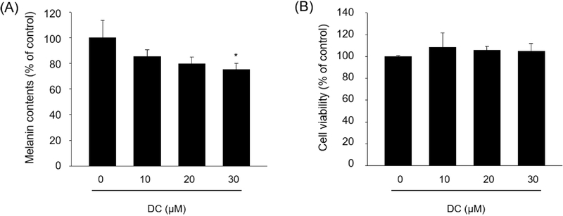
Anti-melanogenic effect of demethoxycurcumin in reconstructed human skin models. (A) Neoderm®-ME tissues were treated with 0–30 µM DC for 4 days, and melanin content was measured at 475 nm after alkaline solubilization. (B) In a separate Neoderm®-ED model, tissues were treated with 0, 10, 20, or 30 µM DC for 3 days, and viability was assessed by the MTT assay. Data are presented as mean ± SD from three independent reconstructed tissue samples (n = 3). For panel A, statistical significance was determined using Student’s t-test versus the untreated control. *p < 0.05 versus the untreated control.
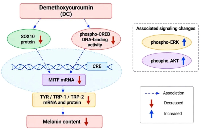
Proposed model of molecular changes associated with the anti-melanogenic effect of demethoxycurcumin. DC treatment was associated with reduced SOX10 abundance and phospho-CREB DNA-binding activity, together with decreased MITF mRNA expression and reduced TYR, TRP-1, and TRP-2 mRNA and protein levels. Increased ERK and AKT phosphorylation is shown as an associated signaling change. The proposed model summarizes these molecular associations and does not establish direct causal relationships among these signaling events.
Collectively, the present findings show that DC treatment is associated with reduced SOX10 abundance and phospho-CREB DNA-binding activity, decreased MITF mRNA expression, and reduced expression of TYR, TRP-1, and TRP-2. PAX3 did not exhibit a consistent concentration-dependent change. Increased ERK and AKT phosphorylation was also observed following DC treatment; however, the causal contribution of these signaling changes to MITF downregulation and melanin suppression remains to be established. Thus, the present study identifies DC as a natural-product-derived anti-melanogenic candidate whose activity is associated with modulation of the SOX10/CREB-MITF regulatory axis. In addition, the Western blot findings were evaluated qualitatively without densitometric quantification from independent experiments. Therefore, the changes in SOX10 abundance and in ERK and AKT phosphorylation should be interpreted as qualitative associations with DC treatment.
Acknowledgments
This research was supported by the Basic Science Research Program through the National Research Foundation of Korea (NRF) funded by the Ministry of Education (RS-2026-25558299) and Bio & Medical Technology Development Program of the National Research Foundation (NRF) funded by the Korean Government (MSIT) (RS-2025-25462906).
Conflicts of Interest
The authors declare that they have no conflicts of interest.
References
-
Hong, S.; Lim, H.; Kim, D.-H. Int. J. Mol. Sci. 2026, 27, 653.
[https://doi.org/10.3390/ijms27020653]

-
Pang, M.; Xu, R.; Xi, R.; Yao, H.; Bao, K.; Peng, R.; Zhi, H.; Zhang, K.; He, R.; Su, Y.; Liu, X.; Ming, D. RSC Med. Chem. 2024, 15, 2226–2253.
[https://doi.org/10.1039/D4MD00224E]
-
Cui, Y.-Z.; Man, X.-Y. Front. Cell Dev. Biol. 2023, 11, 1309557.
[https://doi.org/10.3389/fcell.2023.1309557]
-
Vachtenheim, J.; Borovansky, J. Exp. Dermatol. 2010, 19, 617–627.
[https://doi.org/10.1111/j.1600-0625.2009.01053.x]
-
Wan, P.; Hu, Y.; He, L. Mol. Cell. Biochem. 2011, 354, 241–246.
[https://doi.org/10.1007/s11010-011-0823-4]
-
Saha, B.; Singh, S. K.; Sarkar, C.; Bera, R.; Ratha, J.; Tobin, D. J.; Bhadra, R. Pigment Cell Res. 2006, 19, 595–605.
[https://doi.org/10.1111/j.1600-0749.2006.00348.x]
-
Merecz-Sadowska, A.; Sitarek, P.; Kowalczyk, T.; Zajdel, K.; Kucharska, E.; Zajdel, R. Molecules 2022, 27, 4360.
[https://doi.org/10.3390/molecules27144360]
-
Choi, H.; Yoon, J. H.; Youn, K.; Jun, M. Biomed. Pharmacother. 2022, 147, 112651.
[https://doi.org/10.1016/j.biopha.2022.112651]
-
Fernandes, B.; Cavaco-Paulo, A.; Matamá, T. Biology 2023, 12, 290.
[https://doi.org/10.3390/biology12020290]
-
Jiang, R.; Um, S.; Jeong, H.; Seo, J.; Huh, M.; Kim, Y. R.; Moon, K. Nat. Prod. Sci. 2023, 29, 59–66.
[https://doi.org/10.20307/nps.2023.29.2.59]
-
Feng, D.; Fang, Z.; Zhang, P. Phytomedicine 2022, 107, 154449.
[https://doi.org/10.1016/j.phymed.2022.154449]
-
Qian, W.; Liu, W.; Zhu, D.; Cao, Y.; Tang, A.; Gong, G.; Su, H. Exp. Ther. Med. 2020, 20, 173–185.
[https://doi.org/10.3892/etm.2020.8687]
-
Hatcher, H.; Planalp, R.; Cho, J.; Torti, F. M.; Torti, S. V. Cell. Mol. Life Sci. 2008, 65, 1631–1652.
[https://doi.org/10.1007/s00018-008-7452-4]
- Lee, J. H.; Jang, J. Y.; Park, C.; Kim, B. W.; Choi, Y. H.; Choi, B. T. Int. J. Mol. Med. 2010, 26, 101–106.
-
Tu, C. X.; Lin, M.; Lu, S. S.; Qi, X. Y.; Zhang, R. X.; Zhang, Y. Y. Phytother. Res. 2012, 26, 174–179.
[https://doi.org/10.1002/ptr.3517]
-
Jeon, H.-J.; Kim, K.; Kim, C.; Lee, S.-E. Foods 2023, 12, 926.
[https://doi.org/10.3390/foods12050926]
-
Lv, J.; Yang, Y.; Jia, B.; Li, S.; Zhang, X.; Gao, R. Front. Pharmacol. 2021, 12, 783730.
[https://doi.org/10.3389/fphar.2021.783730]
-
Hong, S.; Lee, S.; Sim, W. J.; Kim, W. C.; Kim, S. Y.; Park, M. H.; Lim, W.; Lim, T. G. J. Funct. Foods 2023, 109, 105813.
[https://doi.org/10.1016/j.jff.2023.105813]
-
Bennett, D. C.; Cooper, P. J.; Hart, I. R. Int. J. Cancer 1987, 39, 414–418.
[https://doi.org/10.1002/ijc.2910390324]
-
Kim, S.-H.; Lee, J.; Jung, J.; Kim, G. H.; Yun, C.-Y.; Jung, S.-H.; Hwang, B. Y.; Hong, J. T.; Han, S.-B.; Jung, J.-K.; Kim, Y. Int. J. Biol. Sci. 2024, 20, 1688–1704.
[https://doi.org/10.7150/ijbs.93120]
-
An, X.; Lv, J.; Wang, F. Eur. J. Pharmacol. 2022, 932, 175231.
[https://doi.org/10.1016/j.ejphar.2022.175231]
-
Jang, J. Y.; Lee, J. H.; Kang, B. W.; Chung, K. T.; Choi, Y. H.; Choi, B. T. Exp. Dermatol. 2009, 18, 232–237.
[https://doi.org/10.1111/j.1600-0625.2008.00794.x]
-
Su, T.-R.; Lin, J.-J.; Tsai, C.-C.; Huang, T.-K.; Yang, Z.-Y.; Wu, M.-O.; Zheng, Y.-Q.; Su, C.-C.; Wu, Y.-J. Int. J. Mol. Sci. 2013, 14, 20443–20458.
[https://doi.org/10.3390/ijms141020443]