
Isolation and Structure Characterization of Aromatic Compounds from Leifsonia shinshuensis and their Antifungal Activities
Abstract
Leifsonia shinshuensis is an endophytic actinomycete and may affect the growth of host plants by producing indole-3-acetic acid (IAA), a phytohormone. However, except for IAA, studies on its secondary metabolites and their potential biological activities remain largely unexplored. In the present study, 11 aromatic compounds (1–11), including a new compound 1, were isolated from L. shinshuensis NPCB2401. Their structures were elucidated by MS, NMR, and ECD data analysis. Biological evaluation revealed that compound 2 exhibited potent antifungal activity against Saccharomyces cerevisiae CEN.PK, with a MIC value of 3.7 μg/mL. These results expanded the structural diversity of metabolites of L. shinshuensis and suggested that L. shinshuensis may contribute to plant defense by producing antifungal metabolites that suppress the growth of fungal pathogens.
Keywords:
Actinomycete, Leifsonia shinshuensis, Antifungal activity, Structure elucidationIntroduction
Actinomycetes are widely distributed in various environments, especially in soil,1 and are well known as a rich source of bioactive secondary metabolites with pharmaceutical potential, such as anticancer,2 antibacterial,3 antifungal,4 and antiviral5 activities. Among these microorganisms, many actinomycetes have also been identified as endophytic bacteria that inhabit internal plant tissues without causing apparent disease symptoms.6 Endophytic bacteria are commonly associated with a wide range of plant species and tissues, including roots, stems, leaves, and seeds, and frequently belong to genera such as Pseudomonas, Bacillus, Burkholderia, Stenotrophomonas, and Micrococcus.7 These microorganisms play essential roles in plant growth and survival by enhancing nutrient acquisition, improving tolerance to environmental stresses, and protecting host plants against phytopathogens.8,9 Such beneficial interactions are often mediated through the production of specialized secondary metabolites, including phytohormones, siderophores, antibiotics, and lipopeptides. These metabolites regulate plant developmental processes and suppress competing microorganisms, thereby contributing to plant fitness and ecological balance.10 Leifsonia shinshuensis is a Gram-positive, aerobic actinomycete that has been characterized as an endophytic bacterium isolated from in vitro cultures of peach rootstocks.11 This bacterium has the ability to produce the phytohormone indole-3-acetic acid (IAA), thereby promoting plant growth.11,12 While the production of IAA by L. shinshuensis has been suggested as a mechanism for plant growth promotion, the full chemical repertoire and the potential ecological roles of its secondary metabolites remain largely unexplored. Considering the diverse biosynthetic potential of actinomycetes, identifying specialized metabolites from this species could provide new insights into the chemical mediation of plant-endophyte symbiosis.
In this study, 11 aromatic compounds, including two isoflavones (1 and 2), a benzofuran (3), three phenylpropanoid/butanoids (4–6), and five indole derivatives (7–11) were isolated from the L. shinshuensis NPCB2401. Their structures were characterized mainly by NMR, MS, and ECD data analysis. The antibacterial and antifungal activities were evaluated for the isolated compounds. Herein, the details of the isolation, structure elucidation, and biological activities are described.
Experimental
General experimental procedures – Nuclear magnetic resonance (NMR) spectra (1H, 13C, COSY, HSQC, and HMBC) were analyzed with a Bruker AVANCE Ⅲ HD 850 NMR equipped with a 5 mm TCI CryoProbe operating at 850 MHz (1H) and 212.5 MHz (13C) or a Bruker AVANCE Ⅲ HD 700 NMR operating at 700 MHz (1H) and 175 MHz (13C), with chemical shifts given in ppm (δ) (Bruker, Karlsruhe, Germany). The HR-ESI-MS data were acquired on an Agilent 6230B time-of-flight mass spectrometer (Agilent Technologies, Santa Clara, CA, USA) coupled to an Agilent 1290 liquid chromatography system and with an OSAKA SODA CAPCELL PAK C18 column (150 mm × 4.6 mm i.d., 5 μm; flow rate: 0.7 mL/min). The LC-MS analysis was conducted on an Agilent 1260 series HPLC system with a diode array detector and a 6130 series ESI mass spectrometer equipped with an analytical Kinetex C18 100 Å column (250 mm × 4.6 mm i.d., 5 μm; flow rate: 0.7 mL/min). Semipreparative high performance liquid chromatography (HPLC) was performed using an Agilent 1260 pump, which was equipped with a Luna C18 100 Å column (250 mm × 10 mm i.d., 10 μm; flow rate: 4.0 mL/min) and Luna Phenyl-Hexyl column (250 mm × 10 mm i.d., 10 μm; flow rate: 4.0 mL/min). Electronic circular dichroism (ECD) spectra were measured on a JASCO J-1500 CD spectrometer (JASCO, Easton, MD, USA). Specific rotations () were measured with a JASCO P-2000 polarimeter (JASCO, Easton, MD, USA).
Bacterial and fungal sources – L. shinshuensis NPCB2401 was isolated from rhizosphere soil samples collected adjacent to trees at Sungkyunkwan University, Suwon, Korea, in January 2024. It was identified as L. shinshuensis (GenBank accession no. NR_043663.1) based on 16S rRNA gene sequence analysis performed by Macrogen (Seoul, Korea), showing 99.3% sequence identity. This strain was deposited in the Laboratory of Natural Product Chemical Biology, School of Pharmacy, Sungkyunkwan University, Korea. Bacillus subtilis 168, Escherichia coli MG1655, and Saccharomyces cerevisiae CEN.PK were provided by Prof. Wonsik Lee (School of Pharmacy, Sungkyunkwan University, South Korea).
Cultivation and small-scale analysis – L. shinshuensis NPCB2401 was incubated for 3 days on Columbia agar plate at 37°C. A single colony was added to 5 mL of Columbia broth (CB) and cultured at 37°C, 250 rpm for 2 days. The bacterial suspension was extracted with 6 mL of ethyl acetate. And the organic layer evaporated to dryness. The dried extracts were solubilized in 200 μL of methanol and analyzed by LC-MS for metabolites profiling. Metabolites analysis was conducted using Phenomenex Luna 5 μm C18 (2) 100 Å (250 mm × 4.6 mm) column with gradient solvent system (0.7 mL/min, 10–100% aqueous acetonitrile, 0.1% formic acid (FA), 20 min).
Extraction and isolation – L. shinshuensis NPCB2401 was cultured in a 14 mL test tube with 5 mL CB for 3 days at 37°C, 250 rpm. Each culture was inoculated into 1 L CB in 2 L Erlenmeyer flasks and incubated for 3 days at 37°C, 250 rpm. A total of 130 L cultivation was conducted and extracted twice with EtOAc in an equal proportion (1:1). The organic layers were evaporated to dryness, yielding 14.5 g of crude extract. The crude extract was fractionated into 30 fractions (Fr. 1–30) via a semipreparative HPLC system (Phenomenex, Luna 10 μm C18 (2) 100 Å (250 mm × 10 mm i.d.) with a gradient elution from 10% to 100% aqueous MeCN with 0.01% TFA over 30 min (flow rate: 4 mL/min). Compound 1 (tR 12.1 min, 0.8 mg) was isolated from fraction 18 using semipreparative HPLC (Phenomenex, Luna 10 μm Phenyl-Hexyl, 250 mm × 10 mm i.d.) with an isocratic system of 45% MeCN with 0.01% TFA (flow rate 4 mL/min for 30 min). Compound 2 (tR 10.5 min, 0.5 mg) was purified from fraction 21 via semipreparative HPLC (Phenomenex, Luna 10 μm Phenyl-Hexyl, 250 mm × 10 mm i.d.) with gradient system of 52–63% MeCN with 0.01% TFA (flow rate 4 mL/min for 30 min). Compound 3 (tR 12.0 min, 0.4 mg) was isolated from fraction 24 using semipreparative HPLC (Phenomenex, Luna 10 μm Phenyl-Hexyl, 250 mm × 10 mm i.d.) with a gradient system of 58-71% MeCN with 0.01% TFA (flow rate 4 mL/min for 30 min). Compounds 4 (tR 8.3 min, 0.5 mg) and 10 (tR 8.2 min, 0.4 mg) were purified using semipreparative HPLC (Phenomenex, Luna 5 μm 250 mm × 10 mm i.d.) with a gradient system of 38–46% MeCN with 0.01% TFA (flow rate 4 mL/min for 30 min). Fraction 17 was isolated by semipreparative HPLC (Phenomenex, Luna 10 μm Phenyl-Hexyl, 250 mm × 10 mm i.d.) with a gradient system of 25–50% MeCN with 0.01% TFA (flow rate 4 mL/min for 30 min) to afford compound 5 (tR 9.9 min, 1.2 mg). Also, compound 6 (tR 12.3 min, 0.6 mg) was isolated from fraction 17 through semipreparative HPLC (Phenomenex, Luna 10 μm Phenyl-Hexyl, 250 mm × 10 mm i.d.) with a gradient system of 39–71% MeCN with 0.01% TFA (flow rate 4 mL/min for 30 min). Compounds 7 (tR 8.4 min, 0.2mg) and 8 (tR 8.5 min, 0.2 mg) were purified using semipreparative HPLC (Phenomenex, Kinetex 5 μm Biphenyl 250 mm × 10 mm i.d.) with an isocratic system of 45% MeCN with 0.01% TFA (flow rate 4 mL/min for 30 min). Compound 9 (tR 12.4 min, 0.5 mg) was isolated from fraction 20 using semipreparative HPLC (Phenomenex, Luna 10 μm Phenyl-Hexyl, 250 mm × 10 mm i.d.) with a gradient system of 45–67% MeCN with 0.01% TFA (flow rate 4 mL/min for 30 min). Compound 11 (tR 16.4 min, 0.4 mg) was isolated from fraction 16 using semipreparative HPLC (Phenomenex, Luna 10 μm Phenyl-Hexyl, 250 mm × 10 mm i.d.) with a gradient system of 25–65% MeCN with 0.01% TFA (flow rate 4 mL/min for 30 min).
Leifsonone (1) – Yellowish gum; −13 (c 0.5, CH3OH); HR-ESI-MS (positive-ion mode) m/z 329.0668 [M + H]+ (calcd for C17H13O7+, 329.0656); UV (MeCN/H2O) λmax (log ε) 220 (2.94), 290 (2.22), 345 (1.80) nm; ECD (c 0.01, MeOH) λmax (Δε) 220 (1.39), 245 (-0.63), 305 (1.48) nm; 1H- and 13C-NMR data, see Table 1.

NMR data of 1 in DMSO-d6a
Irisone B (2) – Yellowish gum; UV (MeCN/H2O) λmax (log ε) 220 (3.21), 270 (2.82), 340 (2.08) nm; 1H-NMR (CDCl3, 700 MHz): δ 12.15 (1H, s, 2′-OH), 8.07 (1H, s, H-2), 7.37 (1H, m, H-4′), 7.17 (1H, dd, J = 7.6, 1.6 Hz, H-6′), 7.11 (1H, dd, J = 8.1, 1.3 Hz, H-3′), 7.01 (1H, td, J = 7.4, 1.3 Hz, H-5′), 6.61 (1H, s, H-10), 6.16 (2H, s, H-8); HR-ESI-MS (positive-ion mode) m/z 299.0575 [M + H]+ (calcd for C16H11O6+, 299.0550).
6-(2-Benzofuranyl)-7-methoxy-1,3-benzodioxol-5-ol (3) – Yellowish gum; UV (MeCN/H2O) λmax (log ε) 220 (3.19), 300 (2.60) nm; 1H-NMR ((CD3)2CO, 700 MHz): δ 7.61 (1H, d, J = 7.7 Hz, H-4′), 7.50 (1H, d, J = 8.1 Hz, H-7′), 7.27 (1H, t, J = 7.6 Hz, H-6′), 7.22 (1H, t, J = 7.4 Hz, H-5′), 6.93 (1H, m, H-3′), 6.34 (1H, s, H-7), 5.98 (2H, s, H-5), 3.95 (3H, s, 3-OCH3); HR-ESI-MS (positive-ion mode) m/z 285.0740 [M + H]+ (calcd. for C16H13O5+, 285.0757).
N-(E)-Feruloyl-3-methoxytyramine (4) – Yellowish gum; UV (MeCN/H2O) λmax (log ε) 220 (3.16), 280 (2.84), 320 (2.88) nm; 1H-NMR (CD3OD, 700 MHz): δ 7.43 (1H, d, J = 15.6 Hz, H-7), 7.12 (1H, d, J = 1.9 Hz, H-6), 7.03 (1H, dd, J = 8.1, 1.9 Hz, H-2), 6.82 (1H, d, J = 1.9 Hz, H-6′), 6.80 (1H, d, J = 8.1 Hz, H-3), 6.72 (1H, d, J = 7.9 Hz, H-3′), 6.67 (1H, dd, J = 8.0, 1.9 Hz, H-2′), 6.41 (1H, d, J = 15.6 Hz, H-8), 3.89 (3H, s, 5-OCH3), 3.83 (3H, s, 5′-OCH3), 3.49 (2H, m, H-8′), 2.77 (2H, t, J = 7.3 Hz, H-7′); HR-ESI-MS (positive-ion mode) m/z 344.1493 [M + H]+ (calcd. for C19H22O5+, 344.1492).
(3E)-4-Phenyl-3-butenoic acid (5) – Yellowish gum; UV (MeCN/H2O) λmax (log ε) 205 (3.53), 250 (3.61) nm; 1H-NMR (CD3OD, 700 MHz): δH 7.38 (2H, d, J = 7.1 Hz, H-2 and H-6), 7.30 (2H, dd, J = 8.4, 7.0 Hz, H-3 and H-5), 7.20 (1H, t, J = 7.3 Hz, H-4), 6.50 (1H, d, J = 15.9 Hz, H-7), 6.34 (1H, dt, J = 15.9, 7.1 Hz, H-8), 3.25 (2H, d, J = 6.8 Hz, H-9); ESI-MS (positive-ion mode) m/z 163.0 [M + H]+ (calcd. for C10H11O2+, 163.0754).
4-Phenylbutanoic acid (6) – Yellowish gum; UV (MeCN/H2O) λmax (log ε) 210 (2.47) nm; 1H-NMR (CD3OD, 700 MHz): δ 7.25 (2H, t, J = 7.6 Hz, H-3 and H-5), 7.19 (2H, m, H-2 and H-6), 7.15 (1H, m, H-4), 2.64 (2H, m, H-7), 2.25 (2H, t, J = 7.4 Hz, H-9), 1.90 (2H, p, J = 7.5 Hz, H-8); ESI-MS (negative-ion mode) m/z 163.1 [M − H]− (calcd. for C10H11O2−, 163.0765).
Indole-3-acetic acid (7) – Yellowish gum; UV (MeCN/H2O) λmax (log ε) 220 (3.18), 275 (2.39) nm; 1H-NMR (CD3OD, 700 MHz): δ 7.57 (1H, d, J = 7.8 Hz, H-4), 7.31 (1H, dt, J = 8.0, 0.9 Hz, H-7), 7.14 (1H, s, H-2), 7.06 (1H, ddd, J = 8.1, 6.8, 1.1 Hz, H-6), 6.98 (1H, ddd, J = 8.0, 6.9, 1.0 Hz, H-5), 3.65 (2H, s, H-10); ESI-MS (positive-ion mode) m/z 176.0 [M + H]+ (calcd. for C10H10NO2+, 176.0706).
Indole-3-ethanol (8) – Yellowish gum; UV (MeCN/H2O) λmax (log ε) 220 (3.02), 275 (2.15) nm; 1H-NMR (CD3OD, 700 MHz): δ 7.53 (1H, d, J = 7.9 Hz, H-4), 7.32 (1H, d, J = 8.0 Hz, H-7), 7.07 (2H, m, H-2 and H-6), 6.99 (1H, t, J = 7.5 Hz, H-5), 3.81 (2H, t, J = 7.3 Hz, H-11), 2.97 (2H, t, J = 7.3 Hz, H-10); ESI-MS (positive-ion mode) m/z 162.1 [M + H]+ (calcd. for C10H12NO+, 162.0913).
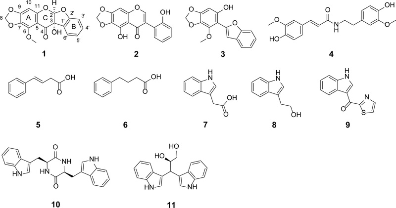
The structures of 1–11 isolated from L. shinshuensis.
Indothiazinone (9) – Yellowish gum; UV (MeCN/H2O) λmax (log ε) 215 (2.96), 275 (2.20), 350 (2.36) nm; 1H-NMR (CD3OD, 700 MHz): δ 9.13 (1H, s, H-2), 8.38 (1H, m, H-4), 8.09 (1H, d, J = 3.1 Hz, H-14), 7.90 (1H, d, J = 3.1 Hz, H-13), 7.50 (1H, m, H-7), 7.28 (2H, ddd, J = 6.9, 4.8, 1.6 Hz, H-5 and H-6); HR-ESI-MS (positive-ion mode) m/z 229.0437 [M + H]+ (calcd. for C12H9N2OS+, 229.0430).
Cyclo-L-Trp-L-Trp (10) – Yellowish gum; −86.3 (c 0.5, CH3OH); UV (MeCN/H2O) λmax (log ε) 220 (3.67), 275 (2.91) nm; 1H-NMR (CD3OD, 700 MHz): δ 7.46 (2H, d, J = 7.9 Hz, H-4 and H-4′), 7.31 (2H, d, J = 8.1 Hz, H-7 and H-7′), 7.10 (2H, ddd, J = 8.1, 6.9, 1.1 Hz, H-6 and H-6′), 7.02 (2H, ddd, J = 8.1, 6.9, 1.0 Hz, H-5 and H-5′), 6.47 (2H, s, H-2 and H-2′), 4.06 (2H, dd, J = 7.1, 3.8 Hz, H-11 and H-11′), 2.93 (2H, m, H-10 and H-10′); HR-ESI-MS (positive-ion mode) m/z 373.1662 [M + H]+ (calcd. for C22H21N4O2+, 373.1659).
Fusarindole B (11) – Brown oil; UV (MeCN/H2O); λmax (log ε) 225 (3.09), 280 (1.79) nm; ECD (c 0.01, CH3OH) λmax (Δε) 220 (3.2), 285 (0.95), 350 (0.3); 1H-NMR (CD3OD, 700 MHz): δ 7.55 (4H, m, H-4 and H-4′), 7.30 (3H, m, H-2, H-7 and H-7′), 7.15 (1H, s, H-2′), 7.03 (2H, overlap, H-6 and H-6′), 6.91 (2H, dt, J = 18.6, 7.5 Hz, H-5 and H-5′), 4.70 (1H, d, J = 6.6 Hz, H-10), 4.49 (1H, td, J = 6.9, 4.0 Hz, H-11), 3.62 (1H, dd, J = 11.1, 4.0 Hz, H-12), 3.50 (1H, td, J = 11.3, 6.6 Hz, H-12); ESI-MS (negative-ion mode) m/z 305.1 [M−H]− (calcd. for C19H17N2O2−, 305.1296).
Antibacterial activity test – B. subtilis 168 and E. coli MG1655 were streaked onto LB agar plates and cultured for 24 h at 37°C. A single colony from each strain was subsequently inoculated into 5 mL of culture broth and incubated for 24 h at 37°C, 250 rpm. Then its OD600 value was adjusted to 0.001. 196 μL of LB medium and 4 μL of each test compound dissolved in DMSO (10 mM) were dispensed into the first column of 96-well plates, and 100 μL of LB broth was added to all other wells. For the DMSO vehicle control, the same volume of DMSO without compound was added to the wells. Serial dilutions were carried out from the second to the last column of the 96-well plates using a multichannel pipette. Then, 100 μL of bacterial culture medium was added to all wells. The plates were incubated at 37°C for 24 h in a shaking incubator and bacterial growth was evaluated by measuring their OD600 value using a microplate reader to determine the survival rate. Each concentration was tested in duplicate wells.
Antifungal activity test – S. cerevisiae CEN.PK was streaked onto a TSB agar plate and cultured for 48 h at 37°C. A single colony was then inoculated into 5 mL of culture broth and incubated for 48 h at 37°C, 250 rpm. Then its OD600 value was adjusted to 0.001. 196 μL of TSB medium and 4 μL of each test compound dissolved in DMSO (10 mM) were dissolved into the first column of a 96-well plate, and 100 μL of TSB broth was added to all other wells. For the DMSO vehicle control, the same volume of DMSO without compound was added to the wells. Serial dilution was carried out from the second to the final column of the 96-well plate using a multichannel pipette. Subsequently, 100 μL of fungal culture medium was added to all wells. The plate was incubated at 37°C for 48 h in a shaking incubator, and fungal growth was evaluated by measuring the OD600 value using a microplate reader to determine the survival rate. Each concentration was tested in duplicate wells.
Results and Discussion
Leifsonone (1) was obtained as a yellowish gum. Its molecular formula was established as C17H12O7 based on the protonated molecular ion observed at m/z 329.0668 [M + H]+ in HR-ESI-MS (calcd for C17H13O7+, 329.0656). UV absorption bands at 220, 290, and 345 nm suggested that 1 is a flavonoid derivative. The 1H-NMR spectrum of 1 exhibited characteristic signals of an ortho-disubstituted benzene ring [δH 7.23 (1H, t, J = 7.7 Hz, H-4′), 7.20 (1H, d, J = 7.6 Hz, H-6′), 6.93 (1H, t, J = 7.5 Hz, H-5′), 6.89 (1H, d, J = 8.1 Hz, H-3′)], an aromatic proton [δH 6.52 (1H, s, H-10)], an oxygenated methine [δH 6.23 (1H, s, H-2)], a methylenedioxy group [δH 6.00 (2H, d, J = 18.3 Hz, H-8)], and a methoxy group [δH 3.77 (3H, s, 6-OCH3)]. The 13C-NMR spectrum of 1 displayed 17 carbon signals, including a ketone [δc 186.8 (C-4)], 12 aromatic carbons [δc 159.7 (C-2′), 154.6 (C-11), 154.3 (C-9), 141.3 (C-6), 133.3 (C-7), 130.9 (C-4′), 127.0 (C-1′), 125.0 (C-6′), 122.1 (C-5′), 110.0 (C-3′), 108.7 (C-5), 94.5 (C-10)], an oxymethine carbon [δc 110.5 (C-2)], a quaternary carbon [δc 81.6 (C-3)], a methylenedioxy group [δc 102.4 (C-8)], and a methoxy group [δc 60.1 (6-OCH3)] (Table 1). The HMBC correlations (Fig. 2), from H-6′ to C-3/C-2′/C-4′ and from H-10 to C-5/C-7, allowed for the assembly of a flavonoid ring system. Furthermore, the HMBC correlations from H-2 to C-2′ in the B ring and to C-11 in the C ring suggested a connection between the B and C rings. Additionally, the HMBC correlations from methylenedioxy protons to C-7/C-9 and from methoxy protons to C-6 confirmed their positions. The absolute configuration of 1 was determined to be 2R and 3S by comparing the experimental ECD spectrum (Fig. 3) with a structurally similar reference compound, (2R,3S)-3,7,4′-trihydroxy-5-methoxycoumaronochromone.13 Therefore, compound 1 was established as a new coumaronochromone-type compound and named leifsonone.
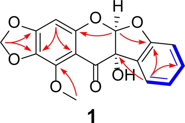
Key COSY (blue bold bonds) and HMBC correlations (red arrows) of 1.
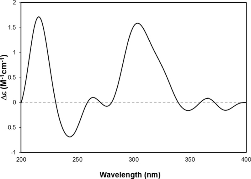
Experimental ECD spectrum of 1.
10 known compounds were identified as irisone B (2),14 6-(2-benzofuranyl)-7-methoxy-1,3-benzodioxol-5-ol (3),15 N-(E)-feruloyl-3-methoxytyramine (4),16 (3E)-4-phenyl-3-butenoic acid (5),17 4-phenylbutanoic acid (6),18 indole-3-acetic acid (7),19 indole-3-ethanol (8),20 indothiazinone (9),21 cyclo-L-Trp-L-Trp (10),22 and fusarindole B (11)23 by comparison of their spectroscopic and spectrometric data with those reported in the literature. Although members of the genus Leifsonia have been studied mainly from taxonomic, ecological, and plant-associated perspectives,24 their secondary metabolites remain relatively underexplored. In particular, chemical investigations of L. shinshuensis have been limited. Thus, the discovery of leifsonone (1) expands the chemical diversity known from this species and highlights L. shinshuensis as an under-explored source of structurally unique aromatic metabolites.
The isolated compounds 1–11 were evaluated for their antibacterial activities against Gram-positive bacteria B. subtilis 168 and Gram-negative bacteria E. coli MG1655, as well as for their antifungal activity against S. cerevisiae CEN.PK, using the broth dilution method. The biological activity results showed that none of the compounds exhibited antibacterial effects, whereas compound 2 revealed potent antifungal activity, with a MIC value of 3.7 μg/mL (Table 2). To the best of our knowledge, this is the first report of the antifungal activity of irisone B (2). This potency is comparable to that of wighteone, a structurally related isoflavone, which reportedly exhibits antifungal activity against S. cerevisiae with a MIC value of 4 μg/mL.25 However, the isoflavone-related scaffold alone does not appear to be sufficient for antifungal activity. In the present assay, leifsonone (1), a structurally related isoflavone-type compound bearing an additional furan ring between the B and C rings, did not show antifungal activity. This observation suggests that the additional furan ring in compound 1 may reduce antifungal activity. Further studies with additional analogues and standard antifungal controls will be required to establish a definitive structure–activity relationship and to more rigorously evaluate the antifungal potency of compound 2.
MIC values of compounds 1–11
In conclusion, our chemical investigation of L. shinshuensis NPCB2401 led to the identification of 11 aromatic metabolites, including a new compound 1. Beyond the potent antifungal activity demonstrated by compound 2 against S. cerevisiae, the presence of indole-3-acetic acid (7, IAA) in the metabolic profile is of particular significance. IAA is one of the most important naturally occurring auxins and plays key roles in plant growth and development, including cell division, elongation, and tissue differentiation. In addition to its role as a plant hormone, IAA is recognized as an important microbial metabolite involved in bacterial physiology, rhizosphere colonization, and plant-microbe interactions.26 Therefore, the ability of this endophytic actinomycete to produce both antifungal agents and growth-promoting hormones like IAA suggests a multifaceted symbiotic relationship with its host plant. Specifically, L. shinshuensis may enhance plant fitness not only by providing a chemical defense shield against fungal pathogens through the production of bioactive aromatics but also by directly modulating host physiological processes via IAA secretion. These findings underscore the potential of L. shinshuensis as a promising biocontrol bacteria and plant growth promoter in sustainable agriculture.
Acknowledgments
This work was supported by the National Research Foundation of Korea (NRF) grant funded by the Korean government (MSIT) (Nos. 2021R1C1C1011045, 2022R1A6A1A03054419) and by the BK21 FOUR Project.
Conflicts of Interest
The authors declare that they have no conflicts of interest.
References
-
Bhatti, A. A.; Haq, S.; Bhat, R. A. Microb. Pathog. 2017, 111, 458–467.
[https://doi.org/10.1016/j.micpath.2017.09.036]

-
Wu, Q.‐B.; Chen, X.‐A.; Lv, Z.‐Y.; Zhang, X.‐Y.; Liu, Y.; Li, Y.‐Q. Appl. Microbiol. Biotechnol. 2021, 105, 4731–4741.
[https://doi.org/10.1007/s00253-021-11344-5]
-
She, W.; Ye, W.; Cheng, A.; Liu, X.; Tang, J.; Lan, Y.; Chen, F.; Qian, P.‐Y. Front. Microbiol. 2021, 12, 635268.
[https://doi.org/10.3389/fmicb.2021.635268]
-
Li, W.; Ding, L.; Li, J.; Wen, H.; Liu, Y.; Tan, S.; Yan, X.; Shi, Y.; Lin, W.; Lin, H.‐W. J. Agric. Food Chem. 2022, 70, 8309–8316.
[https://doi.org/10.1021/acs.jafc.2c02626]
-
Kim, S.‐H.; Ha, T.‐K.‐Q.; Oh, W. K.; Shin, J.; Oh, D.‐C. J. Nat. Prod. 2016, 79, 51–58.
[https://doi.org/10.1021/acs.jnatprod.5b00634]
-
Narsing Rao, M. P.; Lohmaneeratana, K.; Bunyoo, C.; Thamchaipenet, A. Plants 2022, 11, 2976.
[https://doi.org/10.3390/plants11212976]
-
Santoyo, G.; Moreno‐Hagelsieb, G.; del Carmen Orozco‐Mosqueda, M.; Glick, B. R. Microbiol. Res. 2016, 183, 92–99.
[https://doi.org/10.1016/j.micres.2015.11.008]
-
Compant, S.; Duffy, B.; Nowak, J.; Clément, C.; Barka, E. A. Appl. Environ. Microbiol. 2005, 71, 4951–4959.
[https://doi.org/10.1128/AEM.71.9.4951-4959.2005]
-
Lugtenberg, B.; Kamilova, F. Annu. Rev. Microbiol. 2009, 63, 541–556.
[https://doi.org/10.1146/annurev.micro.62.081307.162918]
-
Brader, G.; Compant, S.; Mitter, B.; Trognitz, F.; Sessitsch, A. Curr. Opin. Biotechnol. 2014, 27, 30–37.
[https://doi.org/10.1016/j.copbio.2013.09.012]
-
Liaqat, F.; Eltem, R. 3 Biotech 2016, 6, 120.
[https://doi.org/10.1007/s13205-016-0442-6]
-
Jiang, X.; Li, W.‐W.; Han, M.; Chen, G.; Wu, J.; Lai, S.; Fu, Z.; Zhang, S.; Deng, W.‐W.; Gao, L.; Xia, T. Tree Physiol. 2022, 42, 1043–1058.
[https://doi.org/10.1093/treephys/tpab159]
-
Raksat, A.; Maneerat, W.; Rujanapun, N.; Andersen, R. J.; Pyne, S. G.; Laphookhieo, S. J. Nat. Prod. 2019, 82, 2343–2348.
[https://doi.org/10.1021/acs.jnatprod.9b00216]
- Ayatollahi, S. M.; Moein, M. R.; Kobarfard, F.; Choudhary, M. I. DARU J. Pharm. Sci. 2004, 12, 54–57.
-
McKittrick, B. A.; Stevenson, R. J. Chem. Soc. Perkin 1 1984, 709–712.
[https://doi.org/10.1039/p19840000709]
-
Hwang, J. T.; Kim, Y.; Jang, H.‐J.; Oh, H.‐M.; Lim, C.‐H.; Lee, S. W.; Rho, M.‐C. Molecules 2016, 21, 865.
[https://doi.org/10.3390/molecules21070865]
-
Lee, J. Y.; Lee, J. Y.; Moon, S. S.; Hwang, B. K. J. Agric. Food Chem. 2005, 53, 7696–7700.
[https://doi.org/10.1021/jf050957r]
-
Zhu, R.; Jiang, J.‐L.; Li, X.‐L.; Deng, J.; Fu, Y. ACS Catal. 2017, 7, 7520–7528.
[https://doi.org/10.1021/acscatal.7b01569]
-
Liang, Y.‐S.; Choi, Y. H.; Kim, H. K.; Linthorst, H. J. M.; Verpoorte, R. Phytochemistry 2006, 67, 2503–2511.
[https://doi.org/10.1016/j.phytochem.2006.08.018]
-
Guzmán-López, O.; Trigos, A.; Fernández, F. J.; de Jesús Yañez-Morales, M.; Saucedo-Castaneda, G. World J. Microbiol. Biotechnol. 2007, 23, 1473–1477.
[https://doi.org/10.1007/s11274-007-9392-9]
-
Jansen, R.; Mohr, K. I.; Bernecker, S.; Stadler, M.; Müller, R. J. Nat. Prod. 2014, 77, 1054–1060.
[https://doi.org/10.1021/np500144t]
-
Alqahtani, N.; Porwal, S. K.; James, E. D.; Bis, D. M.; Karty, J. A.; Lane, A. L.; Viswanathan, R. Org. Biomol. Chem. 2015, 13, 7177–7192.
[https://doi.org/10.1039/C5OB00537J]
-
Dai, X.‐M.; Pan, H.‐L.; Lan, W.‐J.; Chen, L.‐P.; Feng, G.‐K.; Deng, R.; Zhu, X.‐F.; Li, H.‐J. Phytochemistry 2022, 204, 113456.
[https://doi.org/10.1016/j.phytochem.2022.113456]
-
Kang, S.‐M.; Asaf, S.; Kim, S.‐J.; Yun, B.‐W.; Lee, I.‐J. J. Biotechnol. 2016, 239, 34–38.
[https://doi.org/10.1016/j.jbiotec.2016.10.004]
-
Yin, H.; Zhao, Y.; Zhang, Y.; Zhang, H.; Xu, L.; Zou, Z.; Yang, W.; Cheng, J.; Zhou, Y. Biotechnol. Lett. 2006, 28, 99–105.
[https://doi.org/10.1007/s10529-005-4954-0]
-
Spaepen, S.; Vanderleyden, J.; Remans, R. FEMS Microbiol. Rev. 2007, 31, 425–448.
[https://doi.org/10.1111/j.1574-6976.2007.00072.x]