
Effects of the Combined Extract of Centella asiatica and Portulaca oleracea on Longitudinal Bone Growth in Adolescent Female Sprague-Dawley Rats
Abstract
This study investigated the effect of FSG-CA, a combined extract of Centella asiatica and Portulaca oleracea, on longitudinal bone growth in female Sprague-Dawley (SD) rats. While previous studies have demonstrated the growth-promoting activity of FSG-CA in males, the potential sex-dependent effects remain unclear. Four-week-old female SD rats received FSG-CA (50 or 100 mg/kg BW/day), vehicle, or recombinant human growth hormone for five weeks. FSG-CA administration significantly enhanced tibial growth plate (GP) height, endochondral bone formation, and chondrocyte proliferation compared with controls. In the liver, FSG-CA increased mRNA expression of insulin-like growth factor-1 (IGF-1) and IGF binding protein-3 (IGFBP-3), while in the GP, IGF-1 and bone morphogenetic protein-2 (BMP-2) expression were upregulated. Serum growth hormone levels were increased. These findings suggest that FSG-CA promotes longitudinal bone growth in females during puberty, potentially via modulation of the GH–IGF-1 axis and local GP signaling pathways.
Keywords:
Centella asiatica, Portulaca oleracea, Longitudinal bone growth, Female, Growth plateIntroduction
Centella asiatica is a perennial herbaceous creeper that is traditionally used as an herbal medicine. It exhibits various pharmacological activities, such as wound healing1,2 and antioxidant3 properties, primarily attributed to its triterpene compounds, including asiaticoside, asiatic acid, and madecassoside.4,5 Portulaca oleracea is a succulent annual plant that has been used as an herbal medicine in many countries.6 It exhibits a wide range of pharmacological effects, including antioxidant7 and anti-inflammatory8 properties, which are attributed to its diverse bioactive constituents, such as flavonoids, alkaloids, fatty acids, terpenoids, and sterols.6
Short stature is defined as a height that falls more than two standard deviations below the mean for a given age, sex, and population group.9 Children with short stature often experience behavioral and psychosocial challenges, including diminished self-esteem, delayed social maturation, or being targets of bullying.10 Individuals with below-average height, even those who do not meet the diagnostic criteria for short stature, have been shown to experience a reduced health-related quality of life.11 Given the recognized role of stature in both physical and psychological development during childhood and adolescence, there is increasing scientific and clinical interest in the management and treatment of this condition.
Recombinant human growth hormone (rhGH), administered subcutaneously, is the standard treatment for short stature caused by growth hormone deficiency.12 However, the clinical effectiveness of rhGH in treating idiopathic short stature, which accounts for approximately 80% of all cases of short stature, remains debated due to the relatively limited gains in final height.13 Concerns also exist regarding long-term use, daily injection pain, inappropriate use in children with normal height, and potential side effects in otherwise healthy individuals.14,15 This has led to increased interest in developing safer and more effective alternatives. In this context, traditional herbal medicines that stimulate longitudinal bone growth are emerging as promising candidates for this purpose.
In a previous in vitro study, we found that C. asiatica extract and P. oleracea extract, respectively, increased insulin-like growth factor-1 (IGF-1) and IGF-binding protein-3 (IGFBP-3) production in hepatocytes and enhanced ALP activity in osteoblasts. Furthermore, their combination exhibited a synergistic effect.16,17 Additionally, our previous in vivo study showed that the combined extract of C. asiatica and P. oleracea (FSG-CA) promoted longitudinal bone growth in adolescent male Sprague-Dawley rats.16
Sex-based differences in responses to natural compounds are well recognized, as males and females may exhibit different pharmacological effects that can influence both efficacy and safety.18,19 Moreover, skeletal growth and maturation are regulated by complex interactions among endocrine, paracrine, and autocrine factors and are profoundly influenced by sex. During puberty, sex hormones play crucial roles in growth plate maturation and longitudinal bone growth, resulting in sex-specific patterns of skeletal development.20,21 Therefore, the biological responses to growth-promoting agents may differ between males and females. Despite the growth-promoting effects of FSG-CA observed in males, its efficacy in females has not yet been investigated. Evaluating FSG-CA in female animals is important not only to determine whether its growth-promoting activity is maintained across sexes but also to identify potential sex-dependent differences in efficacy. Therefore, in the present study, we investigated the effects of oral administration of FSG-CA on longitudinal bone growth in adolescent female Sprague-Dawley rats.
Experimental
Preparation of FSG-CA – FSG-CA, provided by Frombio Co. Ltd. (Yongin, Korea), was prepared according to a previously described method.16,22 Briefly, the C. asiatica extract (CAE) was produced by extracting the dried leaves of C. asiatica with a 30-fold volume of 70% edible ethanol at 50°C for 6 h, followed by filtration, vacuum concentration, and freeze-drying. The P. oleracea extract (POE) was prepared similarly, using the dried whole plant and a 20-fold volume of 70% edible ethanol under the same extraction method. CAE and POE were then blended evenly in a 1:1 ratio to the standardized mixture, used as FSG-CA.
Determination of asiatic acid and adenosine in FSG-CA – Asiatic acid and adenosine were selected as marker compounds for the standardization of FSG-CA because they are representative bioactive constituents of C. asiatica and P. oleracea, respectively, and have been widely used for quality control of these medicinal plants.6,23 The contents of asiatic acid and adenosine in FSG-CA were analyzed by HPLC using a Shimadzu LC-20ADXR system (Shimadzu, Kyoto, Japan), equipped with a Zorbax SB-C18 column (250 × 4.6 mm, 5 μm, Agilent Technologies, Santa Clara, CA, USA) for asiatic acid and a Cadenza CD-C18 column (150 × 4.6 mm, 5 μm, Imtakt, Kyoto, Japan) for adenosine, respectively. The photodiode array detector was set at 210 nm and 260 nm for asiatic acid and adenosine. The mobile phase for analysis of asiatic acid consisted of 0.1% formic acid in water (A) and acetonitrile (B). The gradient elution program was as follows: (A) 55% and (B) 45% at 20 min, (A) 10% and (B) 90% at 30 min, (A) 55% and (B) 45% at 40 min. The mobile phase for analysis of adenosine consisted of 50 mM monobasic potassium phosphate (KH2PO4) in water (A) and methanol (B). The gradient elution program was as follows: (A) 90% and (B) 10% in the 40 min. Both analyses were performed at a flow rate of 1 mL/min and an injection volume of 10 μL. Asiatic acid and adenosine, used as reference standards, were purchased from Tokyo Chemical Industry (Tokyo, Japan) and Sigma-Aldrich (St. Louis, MO, USA), respectively.
Ethical statement and animal care – The animal experimental protocols were approved by the Institutional Animal Care and Use Committee of Hallym University (approved number: Hallym 2023-45) and carried out in compliance with standard guidelines for the care and use of laboratory animals. Female SD rats, three weeks old, were purchased from Dooyeol Biotech Co., Ltd. (Seoul, Korea) and kept in a controlled environment (temperature: 23 ± 3°C; relative humidity: 50 ± 10%; 12 h light/dark cycles), with free access to a commercial rodent diet and tap water.
Experimental design and treatment – After 7-day acclimatization, the rats were randomly divided into four groups: a vehicle-treated group (FCON), a group treated with 50 mg/kg body weight (BW)/day FSG-CA (FF50), a group treated with 100 mg/kg BW/day FSG-CA (FF100), and a group treated with 200 mg/kg BW/day rhGH (FGH). The FF50 and FF100 groups were administered FSG-CA orally once for five weeks, with the doses dissolved in sterile water, whereas the FGH group received daily subcutaneous injections of rhGH at 200 mg/kg BW for five weeks. The animal’s initial and final BW and lengths (from nose to tail, NT) were measured. After the final administration, the rats were anesthetized using 2–3% isoflurane/N2O/O2 mixture, and blood was collected from the orbital vein. Euthanasia was carried out using carbon dioxide asphyxiation, after which the livers, ovaries, and uteri were collected and weighed. The tibiae were also excised, and the soft tissue was removed. They were then measured for both weight and length.
Measurement of serum biochemical indicators – Alanine aminotransferase (ALT) and aspartate aminotransferase (AST) activities in sera were measured using a blood chemistry autoanalyzer (Indiko Plus, Thermo Fisher Scientific, Vantaa, Finland). The levels of estrogen, GH, IGF-1, and IGFBP-3 in serum were measured using specific enzyme-linked immunosorbent assay (ELISA) kits (MyBioSource, San Diego, CA, USA) according to the manufacturer’s instructions.
Measurement of longitudinal bone growth – Tetracycline hydrochloride was utilized as a fluorescent marker to indicate the bone growth line on the tibia surface.24 To evaluate longitudinal bone growth, the rats were given an intraperitoneal injection of tetracycline hydrochloride (20 mg/kg BW, Sigma-Aldrich) 72 h before sacrifice. The dissected tibia was fixed in 4% paraformaldehyde, decalcified using Calci-Clear Rapid (National Diagnostics, Atlanta, GA, USA), embedded in OCT compound (Sakura Finetek, Torrance, CA, USA), and longitudinally sectioned at 30 mm thickness using a cryostat (MICROM HM520, Thermo Scientific, Walldorf, Germany). The sections were then examined under a microscope (AxioImager, Carl Zeiss, Jena, Germany). Longitudinal bone growth was determined by measuring the distance between the fluorescent line and the epiphyseal end line of the growth plate (GP) using Image M1 Software AxioVision 4.8 (Carl Zeiss).
Measurement of epiphyseal growth plate height – The dissected tibia was fixed, decalcified, embedded in paraffin, longitudinally sectioned at 5 μm thickness, and stained with hematoxylin and eosin (H&E, Sigma-Aldrich) according to the manufacturer’s instructions. The stained tissues were observed and photographed under a microscope (AxioImager, Carl Zeiss) in a blind manner. The height of the epiphyseal GP was measured at three different sites using Image M1 Software AxioVision 4.8 (Carl Zeiss).
Immunofluorescence (IF) staining – Sections of paraffin-embedded tibia tissues were deparaffinized, rehydrated, and blocked with 5% bovine serum albumin. IF staining was performed using primary antibodies against IGF-1 and bone morphogenic protein (BMP)-2 (Santa Cruz Biotechnology, Santa Cruz, CA, USA), followed by secondary antibodies conjugated with fluorochrome (Alexa-488 or 564). The nuclei were counterstained with 4′,6-diamidino-2-phenylindole. The stained sections were blindly observed and imaged under a microscope (AxioImager, Carl Zeiss).
Assessment of IGF-1 and IGFBP-3 mRNA expression in the livers – Total RNA from the liver tissue was extracted using TRIzol reagent (Invitrogen Life Technologies, Carlsbad, CA, USA). Single-strand complementary DNA synthesis was performed using a HyperScriptTM RT Master Mix kit (GeneAll Biotechnology, Seoul, Korea). Real-time polymerase chain reaction (PCR) was performed using gene-specific primers as previously reported16 and a QuantiNova SYBR Green PCR kit (Qiagen), as described elsewhere.25 The relative mRNA expression levels of the target genes were normalized to those of glyceraldehyde 3-phosphate dehydrogenase (Gapdh).
Statistical analysis – Statistical analyses were conducted utilizing the Statistical Analysis System (SAS) software, version 9.4 for Windows (SAS Institute, Cary, NC, USA). Data are presented as the mean ± standard error of the mean (SEM). Differences between groups were analyzed by one-way analysis of variance, followed by Duncan’s multiple comparisons test for post-hoc comparisons. A p-value less than 0.05 was considered to indicate a statistically significant difference.
Results and Discussion
C. asiatica extract and P. oleracea extract contain various bioactive compounds. Among the compounds in C. asiatica, asiatic acid—a metabolite of asiaticoside—promotes wound healing by stimulating collagen synthesis and cell proliferation26 and enhances IGF-1 activity.27 Adenosine, one of the bioactive compounds in P. oleracea, facilitates osteoblast differentiation while inhibiting osteoclast activity.28 Based on these properties, asiatic acid and adenosine are considered key bioactive compounds in FSG-CA for their growth-promoting potential. To characterize and standardize FSG-CA, HPLC was performed to quantify the levels of asiatic acid and adenosine. Distinct peaks corresponding to asiatic acid and adenosine were observed at retention times of 13.022 min (detected at 210 nm) and 11.481 min (detected at 260 nm), respectively, matching those of their reference standards. The clear separation of these peaks from adjacent components demonstrated the high specificity of the developed analytical method. Quantitative analysis revealed that FSG-CA contained 13.38 mg/g of asiatic acid and 1.10 mg/g of adenosine (Fig. 1).
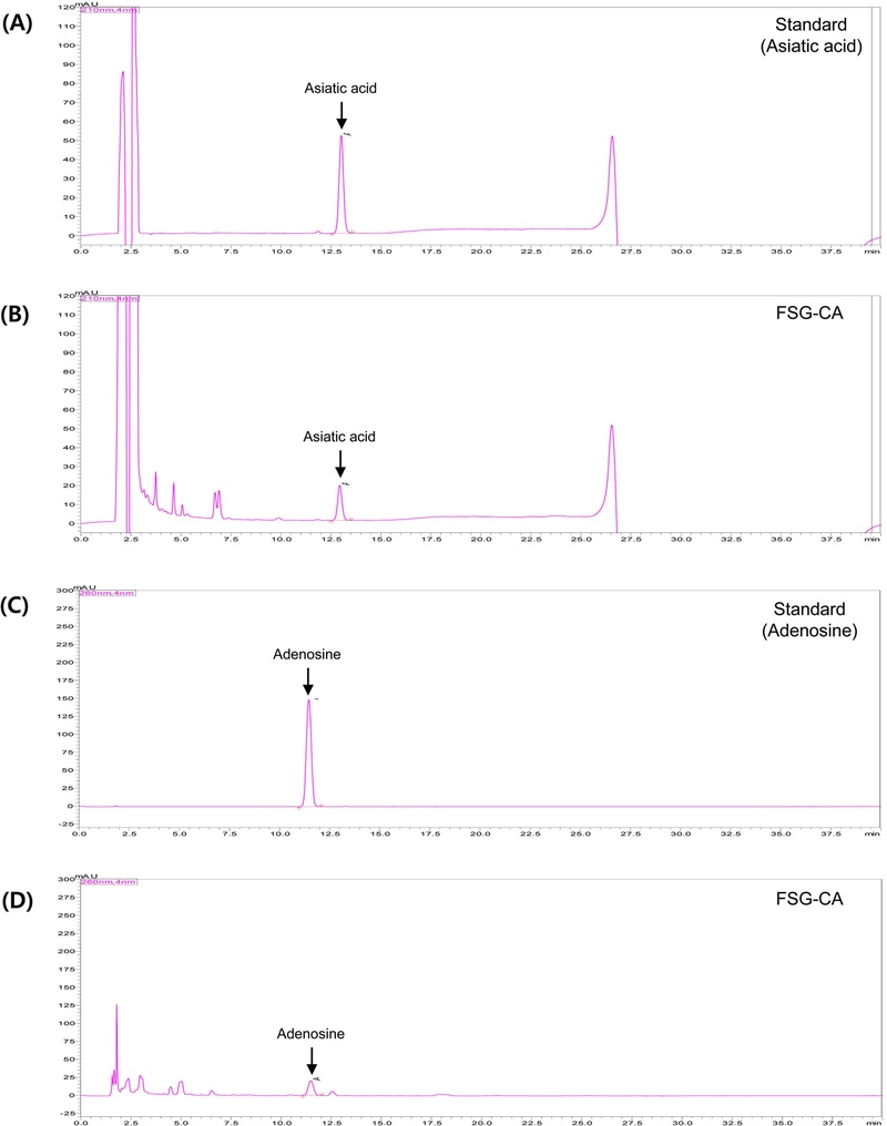
HPLC chromatograms of FSG-CA. (A) Standard, 0.4 mg/mL asiatic acid. (B) 5 mg/mL FSG-CA. (C) standard, 0.5 mg/mL adenosine. (D) 10 mg/mL FSG-CA.
Lee et al.16 previously demonstrated that oral administration of FSG-CA did not cause adverse effects in adolescent male SD rats. To determine whether FSG-CA elicits similar effects in females, we measured BW, liver weight, and serum ALT and AST activities following five weeks of oral FSG-CA administration. No significant changes were observed in BW gain, liver weight, or serum ALT and AST activities in female rats administered FSG-CA (Table 1). Given that natural compounds with growth-promoting properties in females may exert estrogenic or other hormone-like activities that could affect pubertal onset,29 we further examined the impact of FSG-CA on pubertal development by measuring uterine and ovarian weights and serum estrogen levels. FSG-CA administration did not significantly alter the weights of these reproductive organs. Furthermore, FSG-CA administration did not significantly alter serum estrogen levels (Table 1). These findings suggest that FSG-CA is well-tolerated and does not induce observable toxicological effects at doses up to 100 mg/kg BW/day in female SD rats. Nonetheless, additional studies are warranted to fully characterize its safety profile in humans.

Effect of FSG-CA administration on body weight, organ weights, serum ALT and AST activities, and serum estrogen level in adolescent female SD rats.
To examine the effect of FSG-CA on height growth, we measured the initial and final NT lengths and calculated the NT length gain. No significant differences in the initial NT length were observed among the experimental groups. Although the final NT length exhibited an increasing trend following FSG-CA administration, the change did not reach statistical significance. The NT length gain, which represents the difference in NT length before and after FSG-CA administration, was significantly greater in both the FF100 and FGH groups compared to the FCON group. NT length gain increased by 9.2% in the FF100 group and by 16.2% in the FGH group, respectively, compared to the FCON group (Table 2).
Effect of FSG-CA administration on body length and tibia length in adolescent female SD rats.
Among long bones commonly used to assess skeletal growth, including the femur, tibia, and fibula, the tibia is considered the most reliable indicator of stature; therefore, both its length and weight were measured as representative markers of skeletal growth.30 To evaluate the effects of FSG-CA on skeletal growth, tibial weight and length were assessed. Administration of FSG-CA at a dose of 100 mg/kg BW/day, as well as treatment with rhGH at a dose of 200 mg/kg BW/day, significantly increased both the length and weight of the tibia. Compared to the FCON group, tibial length increased by 3.1% and 2.9% in the FF100 and FGH groups, respectively. Similarly, tibial weight increased by 9.8% and 9.2% in the FF100 and FGH groups, respectively (Table 2).
Lee et al.16 reported that administration of FSG-CA at a dose of 100 mg/kg BW/day for 5 weeks in adolescent male SD rats resulted in a 4.8% increase in NT length gain compared to the control group; however, no significant changes were observed in tibial length and weight. These findings suggest that FSG-CA may exert a more pronounced growth-promoting effect on longitudinal bone growth in females than in males.
To evaluate the effect of FSG-CA on endochondral ossification and its longitudinal bone growth, tetracycline fluorescent labeling was employed to bind to newly formed bones within the GP. As shown in Fig. 2A, FSG-CA administration resulted in a significant increase in longitudinal bone growth compared to the FCON group, reaching levels comparable to those observed in the positive control group (FGH). There was no significant difference between the FSG-CA administration of 50 mg/kg BW/day (FF50) and 100 mg/kg BW/day (FF100) (Fig. 2A). These findings indicate that oral administration of FSG-CA at both 50 mg/kg BW/day and 100 mg/kg BW/day promotes longitudinal bone growth to a similar extent as subcutaneous injection of rhGH at 200 μg/kg BW/day in adolescent female rats.
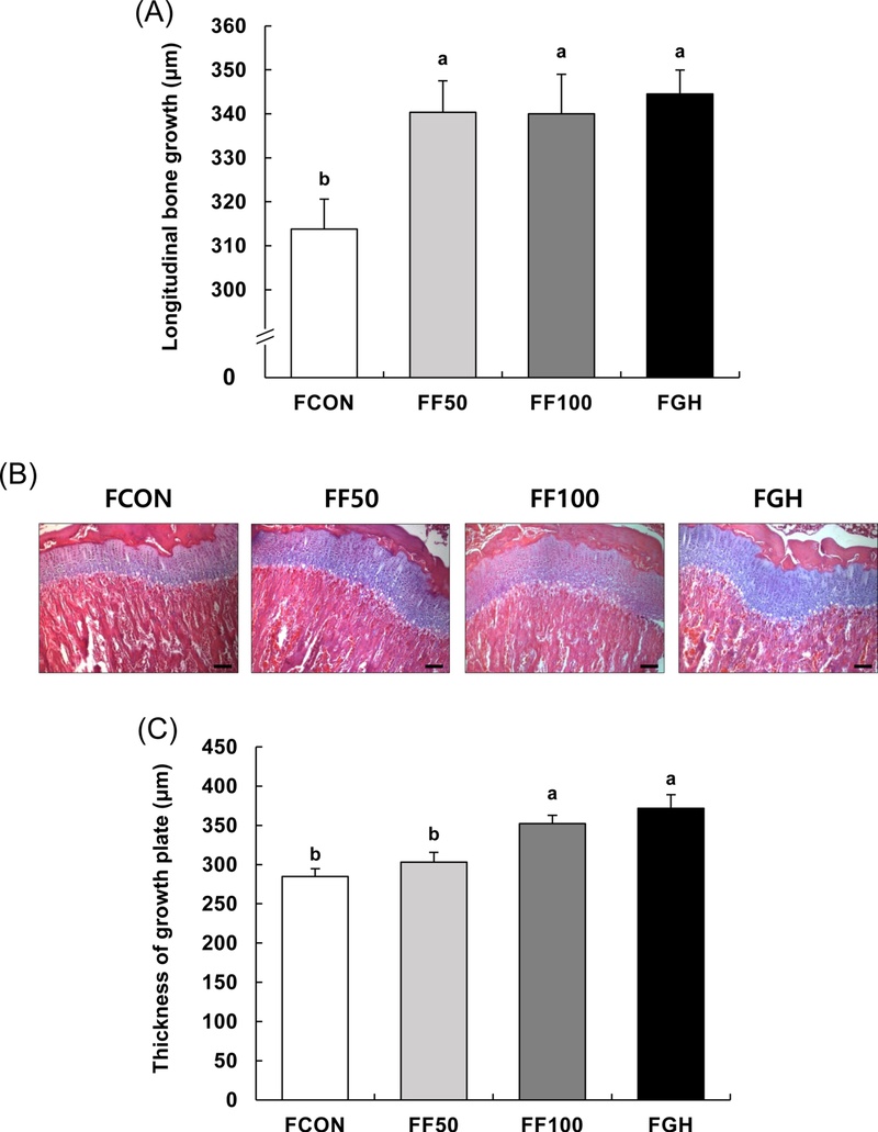
Effect of FSG-CA on longitudinal bone growth in adolescent female SD rats.Four-week-old female SD rats were orally administered FSG-CA (50, 100 mg/kg body weight (BW)/day) or subcutaneously injected with rhGH (200 μg/kg BW/day) for five weeks. (A) The rats were injected intraperitoneally with tetracycline hydrochloride (20 mg/kg BW) 72 h before sacrifice. The tibia section was observed under a fluorescence microscope to measure the longitudinal bone growth. (B) The tibia section was stained with hematoxylin and eosin (H&E). Representative H&E-stained images of tibia tissues (n = 8), 100 x magnification, scale bar = 50 μm. (C) The height of the epiphyseal growth plate was measured. Each bar represents the mean ± SEM (n = 8). Means without a common letter are significantly different at p < 0.05.
The GP is a cartilaginous region located between the metaphysis and epiphysis at the ends of long bones. Within the GP, chondrocytes coordinate longitudinal bone growth through a series of tightly regulated processes, including proliferation, hypertrophy, apoptosis, extracellular matrix production, mineralization, and vascular invasion. The height of the GP serves as an essential morphological indicator of endochondral ossification.31 The height of the proximal tibial GP was measured using H&E staining. Representative histological images of H&E-stained sections are presented in Fig. 2B. Quantitative analysis revealed that the FF50, FF100, and FGH groups exhibited significant increases in GP height by 6.5%, 23.7%, and 30.5%, respectively, compared to the FCON group (Fig. 2C). These results indicate that oral administration of FSG-CA increases growth plate height.
Bone elongation after birth is regulated by a complex interplay between systemic endocrine factors and locally acting paracrine/autocrine factors. Key endocrine factors include GH, IGF-1, thyroid hormones, and sex hormones, with GH recognized as the principal driver of longitudinal bone growth.32 Paracrine/autocrine factors include IGF-1, BMPs, fibroblast growth factor, vascular endothelial growth factor, Wnt proteins, and parathyroid hormone-related protein.33 Bone elongation results from the coordinated interaction between these systemic and local signaling pathways, which together regulate chondrocyte proliferation and hypertrophic differentiation within the GP.33 In particular, the GH–IGF-1 axis plays a central role in promoting longitudinal bone growth.34 This regulation occurs through both endocrine IGF-1 secreted by the liver in response to GH stimulation, and local IGF-1 produced within bone tissue via direct GH action.35
To elucidate the underlying mechanisms contributing to the observed increases in longitudinal bone growth and GP height, serum GH levels were first evaluated. Administration of FSG-CA at dose of 100 mg/kg BW/day led to a significant elevation in serum GH levels, with the FF100 group showing a 45.5% increase compared to the FCON group (Fig. 3A). Given that the liver is the primary source of circulating IGF-1 and its binding protein IGFBP-3 in response to GH stimulation,36 we next examined whether FSG-CA administration upregulates hepatic production of these factors. Quantitative RT-PCR analysis revealed a dose-dependent increase in hepatic IGF-1 mRNA expression following FSG-CA administration. Notably, the FF100 group exhibited approximately a 4-fold increase in IGF-1 mRNA levels relative to the FCON group, reaching levels comparable to those observed in the FGH group (Fig. 3B). In addition, hepatic IGFBP-3 mRNA expression was markedly elevated by FSG-CA administration, showing a 4.5-fold increase in the FF100 group compared to the FCON group (Fig. 3C). Next, serum levels of IGF-1 and IGFBP-3 were analyzed using the relevant ELISA kits. The FF50, FF100, and FGH groups exhibited increases in serum IGF-1 levels by 15.0%, 11.9%, and 8.1%, respectively, compared to the FCON group. However, there was no significant difference in serum IGF-1 levels among the experimental groups (Fig. 3D). These findings indicate that although FSG-CA administration stimulated GH production and enhanced hepatic expression of IGF-1 and IGFBP-3, it did not result in a significant increase in serum IGF-1 or IGFBP-3 levels. When compared with male SD rats, 16 IGFBP-3 showed a similar trend in liver mRNA expression and serum concentration as in males. However, the case of IGF-1 differed from that of males: the increase in IGF-1 mRNA expression in the liver was much greater than in males, whereas the serum concentration was not affected by FSG-CA administration. This suggests that FSG-CA may influence longitudinal bone growth by promoting IGF-1 activity through paracrine or autocrine mechanisms, rather than via endocrine mechanisms mediated by circulating IGF-1.
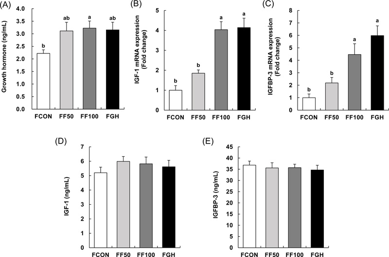
Effect of FSG-CA on growth hormone, IGF-1, and IGFBP-3 expressions in adolescent female SD rats.Four-week-old female SD rats were orally administered FSG-CA (50, 100 mg/kg body weight (BW)/day) or subcutaneously injected with rhGH (200 μg/kg BW/day) for five weeks. (A) Serum growth hormone levels were measured. (B, C) The total RNA in the livers was extracted, reverse-transcribed, and real-time PCR was conducted. The relative mRNA expressions of IGF-1 (B) and IGFBP-3 (C) were analyzed. (D, E) Serum IGF-1 and IGFBP-3 levels were measured. Each bar represents the mean ± SEM (n = 8). Means without a common letter are significantly different at p < 0.05.
IGF-1 plays a pivotal role in promoting longitudinal bone growth.32,33 Evidence suggests that locally synthesized IGF-1 may have a more critical function than GH in this process, as IGF-1-deficient mutant models exhibit significantly more severe growth impairments compared to those with disrupted GH function.37 In vitro studies demonstrated that FSG-CA enhances osteogenic activity in MC3T3-E1 osteoblastic cells.22 Furthermore, in adolescent male SD rats, FSG-CA administration led to increased IGF-1 expression in the proximal tibia.16 These findings indicate that bone-derived IGF-1 contributes substantially to longitudinal bone growth, potentially mediated by FSG-CA administration. To further investigate this mechanism, IGF-1 protein expression was assessed in the proximal tibial GP via immunofluorescence staining. Both FSG-CA and rhGH administration significantly upregulated IGF-1 expression in the GP relative to the FCON group (Fig. 4).
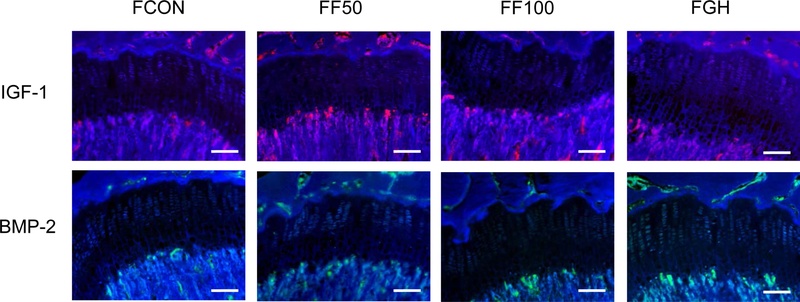
Effect of FSG-CA on IGF-1 and BMP-2 expressions in the tibia of adolescent female SD rats.Four-week-old female SD rats were orally administered FSG-CA (50, 100 mg/kg body weight (BW)/day) or subcutaneously injected with rhGH (200 μg/kg BW/day) for five weeks. The tibia section was stained with IGF-1 and BMP-2 antibodies. Representative staining images are shown (n = 5), 200 × magnification, scale bar = 50 μm.
We investigated the expression of BMP-2 along with IGF-1 in proximal tibial GP, given that BMPs are members of the transforming growth factor-β (TGF-β) superfamily and act as both growth and differentiation factors. Among the various BMP isoforms, BMP-2 has been shown to play a key role in the development of the GP and in promoting longitudinal bone growth by stimulating the proliferation and hypertrophy of chondrocytes within the GP.38 BMP-2 protein expression in the proximal tibial GP was evaluated by immunofluorescence staining, revealing that administration of FSG-CA increased BMP-2 expression compared to the FCON group (Fig. 4). This finding suggests that FSG-CA administration enhances endochondral ossification by upregulating BMP-2 expression, thereby contributing to longitudinal bone growth.
In conclusion, the administration of FSG-CA increased endochondral bone growth and GP height in adolescent female SD rats. FSG-CA also increased the mRNA expression of IGF-1 and IGFBP-3 in the liver. However, it did not affect circulating IGF-1 and IGFBP-3 concentrations. Considering immunohistochemical studies, the effect of FSG-CA on longitudinal bone growth appears to be due to an increase in local IGF-1 and BMP-2 expression at the GP. Furthermore, this seems to be mediated through the GH-IGF-1 axis, predominantly via local paracrine/autocrine mechanisms rather than systemic endocrine regulation. These results suggest that FSG-CA administration during puberty in female SD rats can increase longitudinal bone growth via the GH-IGF-1 pathway.
Acknowledgments
The authors thank the Frombio Co., Ltd. for providing the mixture of Centella asiatica extract and Portulaca oleraceae extract (FSG-CA).
Conflict of Interest
The authors declare that they have no conflicts of interest.
References
-
Brinkhaus, B.; Lindner, M.; Schuppan, D.; Hahn, E. G. Phytomedicine 2000, 7, 427–448.
[https://doi.org/10.1016/S0944-7113(00)80065-3]

- Sunilkumar; Parameshwaraiah, S.; Shivakumar, H. G. Indian J. Exp. Biol. 1998, 36, 569–572.
-
Jayashree, G.; Kurup Muraleedhara, G.; Sudarslal, S.; Jacob, V. B. Fitoterapia 2003, 74, 431–434.
[https://doi.org/10.1016/S0367-326X(03)00121-7]
-
Gohil, K. J.; Patel, J. A.; Gajjar, A. K. K. Indian J. Pharm. Sci. 2010, 72, 546–556.
[https://doi.org/10.4103/0250-474X.78519]
-
Sun, B.; Wu, L.; Wu, Y.; Zhang, C.; Qin, L.; Hayashi, M.; Kudo, M.; Gao, M.; Liu, T. Front. Pharmacol. 2020, 11, 568032.
[https://doi.org/10.3389/fphar.2020.568032]
-
Zhou, Y.‐X.; Xin, H.‐L.; Rahman, K.; Wang, S.‐J.; Peng, C.; Zhang, H. Biomed. Res. Int. 2015, 2015, 925631.
[https://doi.org/10.1155/2015/925631]
-
Lim. Y. Y.; Quah, E. P. L. Food Chem. 2007, 103, 734–740.
[https://doi.org/10.1016/j.foodchem.2006.09.025]
-
Hwang, J. T.; Kim, Y.; Jang, H.‐J.; Oh, H.‐M.; Lim, C.‐H.; Lee, S. W.; Rho, M.‐C. Molecules 2016, 21, 865.
[https://doi.org/10.3390/molecules21070865]
-
Cohen, P.; Rogol, A. D.; Deal, C. L.; Saenger, P.; Reiter, E. O.; Ross, J. L.; Chernausek, S. D.; Savage, M. O.; Wit, J. M.; 2007 ISS Consensus Workshop participants, J. Clin. Endocrinol. Metab. 2008, 93, 4210–4217.
[https://doi.org/10.1210/jc.2008-0509]
-
Voss, L. D.; Sandberg, D. E. Eur. J. Endocrinol. 2004, 151, S29–S33.
[https://doi.org/10.1530/eje.0.151s029]
-
Christensen, T. L.; Djurhuus, C. B.; Clayton, P.; Christiansen, J. S. Clin. Endocrinol. 2007, 67, 407–412.
[https://doi.org/10.1111/j.1365-2265.2007.02901.x]
-
Wit, J. M.; Kamp, G. A.; Rikken, B. Pediatr. Res. 1996, 39, 295–302.
[https://doi.org/10.1203/00006450-199602000-00018]
- Bryant, J.; Baxter, L.; Cave, C. B.; Milne, R. Cochrane Database Syst. Rev. 2007, CD004440.
-
Hintz, R. L. BMJ 2004, 328, 907–908.
[https://doi.org/10.1136/bmj.328.7445.907]
-
Silverman, B. L.; Blethen, S. L.; Reiter, E. O.; Attie, K. M.; Neuwirth, R. B.; Ford, K. M. J. Pediatr. Endocrinol. Metab. 2002, 15, 715–722.
[https://doi.org/10.1515/JPEM.2002.15.S2.715]
-
Lee, H. S.; Jung, J. I.; Lim, S.; Jang, Y.; Kim, E. J. Food Nutr. Res. 2025, 13, 232–239.
[https://doi.org/10.12691/jfnr-13-7-1]
- Shim, T. J.; Kim, J. H.; Hong, I. K.; Jang, Y. S.; Min, S. Y.; Kim, E. R., Kim, H. B.; Kim, J. I.; Lee, E. J.; Park, C. W.; Kim, E. J.; Jung J. I. Korean Intellectual Property Office, 10–2700325.
-
Beery, A. K.; Zucker, I. Neurosci. Biobehav. Rev. 2011, 35, 565–572.
[https://doi.org/10.1016/j.neubiorev.2010.07.002]
-
Soldin, O. P.; Mattison, D. R. Clin. Pharmacokinet. 2009, 48, 143–157.
[https://doi.org/10.2165/00003088-200948030-00001]
-
Emons, J.; Chagin, A. S.; Sävendahl, L.; Karperien, M.; Wit, J. M. Horm. Res. Paediatr. 2011, 75, 383–391.
[https://doi.org/10.1159/000327788]
-
Weise, M.; De‐Levi, S.; Barnes, K. M.; Gafni, R. I.; Abad, V.; Baron, J. Proc. Natl. Acad. Sci. U S A 2001, 98, 6871–6876.
[https://doi.org/10.1073/pnas.121180498]
-
Lee, H. S.; Jung, J. I.; Lim, S.; Jang, Y.; Kim, E. J. J. Nutr. Health. 2025, 58, 253–262.
[https://doi.org/10.4163/jnh.2025.58.3.253]
-
Gray, N. E.; Alcazar Magana, A.; Lak, P.; Wright, K. M.; Quinn, J.; Stevens, J. F.; Maier, C. S.; Soumyanath, A. Phytochem. Rev. 2018, 17, 161–194.
[https://doi.org/10.1007/s11101-017-9528-y]
-
Pautke, C.; Vogt, S.; Kreutzer, K.; Haczek, C.; Wexel, G.; Kolk, A.; Imhoff, A. B.; Zitzelsberger, H.; Milz, S.; Tischer, T. J. Anat. 2010, 217, 76–82.
[https://doi.org/10.1111/j.1469-7580.2010.01237.x]
-
Lim, S.‐M.; Lee, H. S.; Jung, J. I.; Kim, S. M.; Kim, N. Y.; Seo, T. S.; Bae, J.‐S.; Kim, E. J. Nutrients 2019, 11, 1190.
[https://doi.org/10.3390/nu11051190]
-
Diniz, L. R. L.; Calado, L. L.; Duarte, A. B. S.; de Sousa, D. P. Metabolites 2023, 13, 276.
[https://doi.org/10.3390/metabo13020276]
-
Ariani, A.; Ghofar, I. F.; Khotimah, H.; Nurdiana, N.; Rahayu, M. Open Vet. J. 2023, 13, 629–637.
[https://doi.org/10.5455/OVJ.2023.v13.i5.16]
-
Mediero, A.; Cronstein, B. N. Trends Endocrinol. Metab. 2013, 24, 290–300.
[https://doi.org/10.1016/j.tem.2013.02.001]
-
Jefferson, W. N.; Patisaul, H. B.; Williams, C. J. Reproduction 2012, 143, 247–260.
[https://doi.org/10.1530/REP-11-0369]
-
Kim, J. Y.; Song, M.; Lee, D.; Song, J.; Park, S. W.; Park, J.; Park, S.; Choi, H.‐Y.; Kim, H. Molecules 2013, 18, 13271–13282.
[https://doi.org/10.3390/molecules181113271]
- Lee, D.; Lee, S. H.; Lee, Y. H.; Song, J.; Kim, H. Evid. Based. Complement. Alternat. Med. 2017, 2017, 6935802.
-
Lui, J. C.; Nilsson, O.; Baron, J. J. Mol. Endocrinol. 2014, 53, T1–9.
[https://doi.org/10.1530/JME-14-0022]
-
Racine, H. L.; Serrat, M. A. Curr. Osteoporos. Rep. 2020, 18, 210–227.
[https://doi.org/10.1007/s11914-020-00570-x]
-
Blair, J. C.; Savage, M. O. Trends Endocrinol. Metab. 2002, 13, 325–330.
[https://doi.org/10.1016/S1043-2760(02)00631-8]
-
Yakar, S.; Liu, J. L.; Stannard, B.; Butler, A.; Accili, D.; Sauer, B.; LeRoith, D. Proc. Natl. Acad. Sci. U S A 1999, 96, 7324–7329.
[https://doi.org/10.1073/pnas.96.13.7324]
-
Yakar, S.; Rosen, C. J.; Beamer, W. G.; Ackert‐Bicknell, C. L.; Wu, Y.; Liu, J.‐L.; Ooi, G. T.; Setser, J.; Frystyk, J.; Boisclair, Y. R.; LeRoith, D. J. Clin. Invest. 2002, 110, 771–781.
[https://doi.org/10.1172/JCI200215463]
-
Baker, J.; Liu, J. P.; Robertson, E. J.; Efstratiadis, A. Cell 1993, 75, 73–82.
[https://doi.org/10.1016/S0092-8674(05)80085-6]
-
Lee, D.; Kim, Y.-S.; Song, J.; Kim, H. S.; Lee, H. J.; Guo, H.; Kim, H. Molecules 2016, 21, 461.
[https://doi.org/10.3390/molecules21040461]