Antioxidant Activity of Vitex rotundifolia Seeds and Phytochemical Analysis Using HPLC-PDA
Abstract
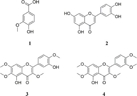
This study assessed in vitro antioxidant activity (ABTS+ and DPPH) of Vitex rotundifolia seeds collected from two different regions in Korea (Jungjang City and Sindu City). Three extraction methods using ethanol, methanol, and water were prepared separately and subjected to quantification by reverse-phase high-performance liquid chromatography-photodiode array (HPLC-PDA) analysis as well as antioxidant testing. Among them, the water-based extract exhibited superior activity in the ABTS+ compared with the ethanol- and methanol-based extracts, while the DPPH assay analysis, revealed that the methanol-based extract had very low antioxidant activity. The concentrations of vanillic acid (1), luteolin (2), vitexicarpin (3), and artemetin (4) were quantified using HPLC-PDA analysis. Vanillic acid (1) was identified as the main antioxidant in V. rotundifolia seeds. Combining the antioxidant activity and quantitative analysis results, the water-based extract was considered to have the highest antioxidant activity. Furthermore, vanillic acid (1) was detected in the leaves and stems of V. rotundifolia plants from different regions, indicating that this species has the potential for use in future antioxidant-applications.
Keywords:
Vitex rotundifolia, Antioxidant activity, HPLC, Quantitative analysis, Vanillic acidIntroduction
Vitex rotundifolia (VR), a species of the family Verbenaceae, is a halophytic deciduous broad-leaved shrub that inhabits coastal areas or islands in the Pacific.1 The fruit is a drupe and ripens to dark purple in October to November.2 The leaves and stems have dense hairs, and the roots develop from the stems. Other representative halophytes include Salicornia europaea, Artemisia capillaris, Glehnia littoralis, and Carex kobomugi.3 Because VR plants on the coast are exposed directly or indirectly to salt from seawater, they accumulate anti-stress compounds, such as flavonoids, to survive the harsh environment. Therefore, it is expected that the secondary metabolites in coastal VR populations will differ from those inhabiting inland areas.4,5
VR contains various bioactive constituents with antioxidant, anti-inflammatory, anti-depressant, and sedative effects, including flavonoids and terpenoids, and these are distributed throughout all parts of the plant.6-8 The seeds of VR, which are called “Manhyeongja” in Oriental medicine, also have a unique scent and have the effect of relieving headaches, so they have traditionally been used as a pillow material.1,9 In addition, the leaves and stems of VR have been used as a desiccant to remove moisture indoors and as a bathing material for their positive effects on the nervous system.10 VR also contains about 76 essential oil components that have various biologically active effects, including monoterpenes, phenol compounds, and flavonoids.11-13 In the search for physiologically active compounds, chemical content analysis and activity tests have been conducted on VR populations in different regions.14 In Korea, VR seeds are used as a filling material for pillows, while residents of Jeju Island, Chungnam Province, and the west coast and island regions of Gyeonggi Province believe that boiling them in water and drinking it alleviates headaches.15
Reactive oxygen species (ROS) are produced in the body via the metabolism of oxygen. Though they play critical roles in the regulation of various cell functions, at excessive levels they can damage cell structures, tissue, and protein, leading to oxidative stress, disease, and aging.16-18 To lower the concentration of free radicals in the body, it is desirable to consume foods containing antioxidants. 2,2-Diphenyl-1-picrylhydrazyl (DPPH) is a stable water-soluble free radical with a maximum absorbance of 510 to 530 nm19-21 that is widely employed to assess radical scavenging activity. 2,2'-azino-bis(3-ethylbenzothiazoline-6-sulfonic acid) (ABTS) assays have recently been developed for the same purpose and have been used for screening plant extracts and biological fluids.22 DPPH assays are also used to measure the antioxidant activity of the phenolic structures and aromatic amine components of natural products.23,24
In this study, we aimed to determine and quantify the phytochemical compounds in ethanol (EtOH), methanol (MeOH), and distilled water (DW) extracts of VR collected from different regions in Korea using high-performance liquid chromatography-photodiode array (HPLC-PDA) analysis. In addition, the in vitro antioxidant activity of the extracts was analyzed using the ABTS and DPPH assays.
Experimental
Plant materials – VR seeds were collected from the cities of Jungjang and Sindu in Chungcheong Province, Korea. Additionally, VR leaves and stems collected from Jeju Island and Chungnam Province were purchased from the Korea Plant Extract Bank (Korea Research Institute of Bioscience and Biotechnology, Korea).
Instruments, chemicals, and reagents – Quantitative analysis was conducted using an HPLC system (Waters Alliance e2695 Separations Module, USA) equipped with an auto-sampler, a pump, and a PDA (Waters 2998 PDA detector, USA). HPLC-grade solvents water, acetic acid (ACC), and acetonitrile (ACN) were purchased from J. T. Baker (Phillipsburg, PA, USA). EtOH, MeOH, and DW purchased from J. T. Baker (Phillipsburg, PA, USA) were used for the analysis. An Epoch microplate spectrophotometer (BioTek, Winooski, VT, USA) was employed for microplate reader analysis. Compounds 1–4 (vanillic acid, luteolin, vitexicarpin, and artemetin, respectively) were obtained from Natural Product Institute of Science and Technology (www.nist.re.kr), Anseong, Korea (Fig. 1).
Extraction methods and preparation of the samples – Dried VR seeds were pulverized and used for extraction. Each sample (5 g) was extracted three times with EtOH, MeOH, and DW (80°C, 100 mL × 3 h) and concentrated in vacuo (50°C). Each extract (50 mg) was dissolved in 1 mL of 70% MeOH and filtered through a syringe filter (0.45 μm) to prepare the sample stock solutions.
ABTS+ radical scavenging assays – ABTS+ and potassium persulfate were dissolved in DW (pH 7.4) to 7.4 and 2.6 mM, respectively, and the solutions were left in a dark place at 4°C for 24 h. Following this, 10 μL of each sample was added to 200 μL of an ABTS+ stock solution prepared by diluting it with distilled water so that the absorbance was 1.00 ± 0.2 at 734 nm. After sitting in the dark for 30 min, the concentration of the residual radicals was measured at 734 nm using a microplate reader (Epoch, BioTek, Winooski, VT, USA). Ascorbic acid at a concentration range of 0.0–0.2 mg/mL was used as a positive control. The results were expressed as the concentration of the sample needed for the 50% inhibition (IC50) of the ABTS+ radicals.
[ABTS radical scavenging activity (%) = (Blank O.D – Sample O.D) / Blank O.D × 100]
DPPH radical-scavenging assays – DPPH (200 μL, 0.2 mM; Sigma, USA) was dissolved in 95% EtOH, and 10 μL of each extract was added to this. After the mixture was allowed to react in the dark for 30 min, the absorbance of the residual radicals was measured at 514 nm with a microplate reader (Epoch, BioTek). Ascorbic acid was used as a positive control. The results were reported as the IC50 values.
[DPPH radical scavenging activity (%) = (Blank O.D – Sample O.D) / Blank O.D × 100]
HPLC-PDA conditions – HPLC analysis was conducted using a reverse-phase HPLC system with a reverse-phase INNO-C18 column (25 cm × 4.6 mm, 5 μm). The detection wavelength and injection volume were 280 nm and 10 μL, respectively. The auto-sampler was set at room temperature. A mobile phase of 0.5% ACC in DW (A) and 0.5% ACC in ACN (B) was employed with gradient elution at a flow rate of 0.9 mL/min. The gradient elution system was programmed as follows: 90% A (0–5 min); 90% A → 52% A (5–40 min); 52% A (40–45 min), 52% A → 0% A (45–50 min); 0% A (50–53 min); 0% A → 90% A (53–55 min); 90% A (55–60 min).
Calibration curves – Five different concentrations of the standard compound were prepared. The calibration curve was constructed by plotting the peak area of each solution against its corresponding concentration. Linearity was then determined based on the correlation coefficient (r2). The content of the analyte was measured on the corresponding calibration curve. The standard calibration function was calculated using the peak area (y) and concentration (x, mg/mL), with the mean ± standard deviation (n = 5) presented (Table 1).
Results and Discussion
In order to compare the antioxidant capabilities of VR seeds collected from different regions, the in vitro antioxidant activity against ABTS+ and DPPH for the compounds extracted from the VR seeds using three solvents (EtOH, MeOH, and DW) was assayed (Table 2). In the ABTS+ assay, the highest antioxidant activity was exhibited by the DW-based extract from VR seeds from Jungjang City (IC50, 0.67 mg/mL) and Sindu City (IC50, 0.84 mg/mL). In the DPPH assay, the EtOH-based extract from VR seeds from Jungjang City (IC50, 2.35 mg/mL) and the DW-based extract from VR seeds from Sindu City (IC50, 2.67 mg/mL) had the highest activity. These assays confirmed that the extraction efficiency of DW tended to be higher than that of the two tested alcohols, and the VR seeds from Jungjang City exhibited the highest antioxidant activity. Based on these results, when the VR seeds are used as antioxidants, it is better to extract compounds from them using DW. It was also confirmed that VR seeds are useful natural antioxidants because they demonstrated excellent electron-donating ability. These seeds contain high levels of natural polyphenols and flavonoids, while their polyphenol content is reported to be higher than that of medicinal plants used as Oriental herbal medicines.25
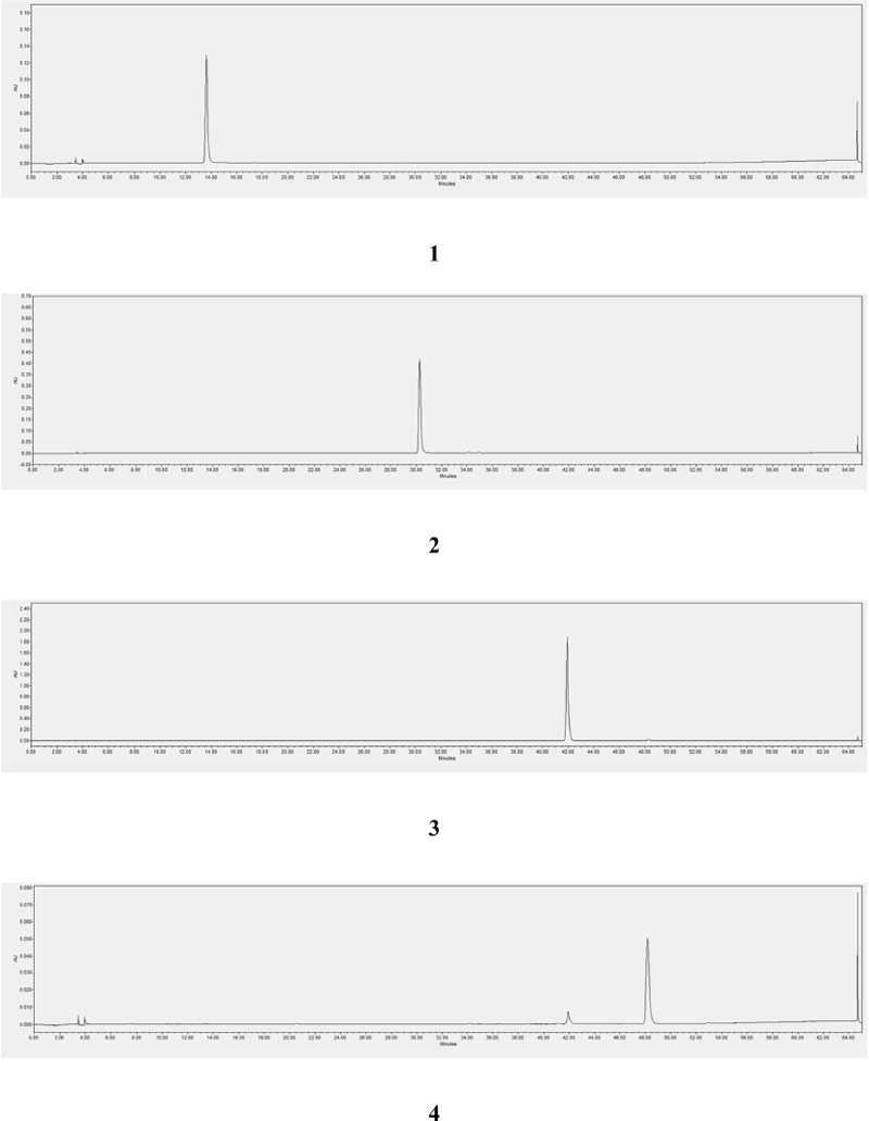
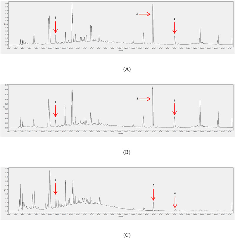
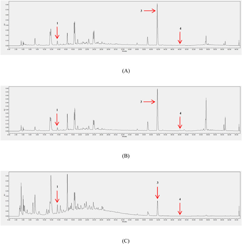
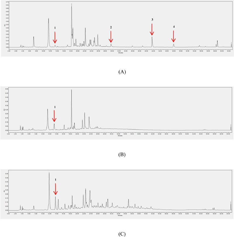
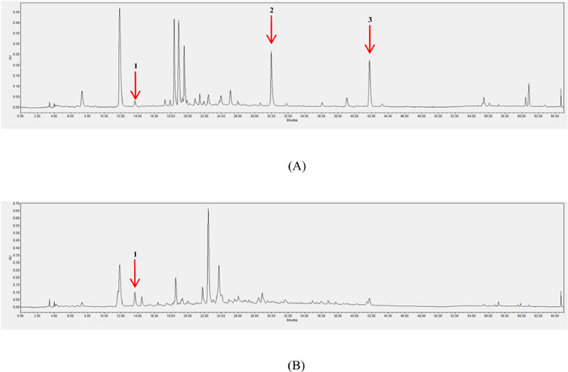
In order to quantify the compounds contained in VR seeds, the extracts were analyzed for compounds 1–4 using HPLC-PDA (Fig. 2). Good linearity was obtained within the tested concentration range, with a correlation coefficient (r2) of 0.9989–1.0000 (Table 1). The flavonoids and phenolic acids of VR seeds were used as marker compounds to evaluate their pharmacological value. It has been reported that these compounds are primarily responsible for bioactivity.26 The results of the quantitative analysis revealed that the flavonoid and phenolic acid content varied depending on the solvent used for extraction (Table 3). Compound 1 is known to have high antioxidant activity, so it has been frequently targeted in the development of functional foods and pharmaceuticals.27 Compounds 1, 3, and 4 were present in all of the VR seed extracts, but compound 2 was not (Figs. 3 and 4). Compound 1 was the main component in the EtOH-based extract from VR seeds from Jungjang City (1.31 mg/g ext.; Table 3), and this extract also exhibited the strongest activity in the DPPH assays (Table 2). In addition, the highest compound 1 concentration in the VR seeds from Sindu City was observed in the DW-based extract (0.89 mg/g ext.; Table 3), which was in accordance with the results showing that this extract had the highest antioxidant activity in the ABTS+ assays. The concentrations of compounds 3 and 4 differed significantly depending on the solvent used for extraction and the region from which the seeds were collected, thus the extraction method should be selected in accordance with the intended application purpose. Compounds 1 and 2 have demonstrated potent antioxidant activity in previous studies, while the antioxidant activity of compounds 3 and 4 have been less well-studied.7,28 However, compounds 3 and 4 have been reported to have the potential to effectively inhibit peroxynitrite formation from nitric oxide (NO) and superoxide ions (O2–), and thus may have high antioxidant activity.29
Though the leaves and stems of VR are less widely utilized than the seeds in Korea, content analysis and assessment of the antioxidant activity of their extracts were conducted to determine their potential for use in various applications. Compound 1 was detected in the MeOH-based extracts for leaves and stems from Jeju Island and Chungnam Province (Figs. 5 and 6), while compound 2, which was not detected in the VR seeds, was detected in VR leaves at concentrations of 1.29 and 8.39 mg/g ext. in these regions, respectively (Table 4). Compound 3 was detected in the leaves from both regions but not in the stems, while compound 4 was detected only in the leaves (6.80 mg/g ext.) from Jeju Island. Although compound 1 was detected in all parts of the VR plants from both regions, compounds 2–4 were not detected in the stems from any region. A previous study reported that compounds 2–4 suppressed anti-apoptotic expression in human cancer cells and thus exhibited activity that promoted the death of cancer cells, so it is considered beneficial to use the leaves of VR for their anti-cancer effects.30
In the precent study, the in vitro antioxidant activity of VR seeds, which are a potential resource for the food and pharmaceutical industries, was identified according to the solvent used for extraction. The main phytochemical compounds contributing to this antioxidant activity were also identified, and the differences in the concentrations of these compounds between regions, parts of the plant and extraction solvents were determined. It was found that the most appropriate extraction method depends on the application, and the relationship between the antioxidant activity of VR seed extracts and the constituent compounds was verified. The DW-based extract exhibited antioxidant activity that was superior to that of the extracts produced using other solvents, and compound 1 was detected in all samples, suggesting that it is primarily responsible for the antioxidant activity. This study thus provides basic data for the future utilization of VR, which has been underutilized to date, by analyzing the characteristics and concentrations of the active compounds and highlights its potential value as a resource for food and medicinal applications.
Acknowledgments
This research was supported by the Studies on the development of methods for the application of native plant resources based on traditional ethnobotanical knowledge in Korea (KNA1-2-34, 17-9) program, Korea National Arboretum, Pocheon, Republic of Korea.
Conflicts of Interest
The authors declare that they have no conflicts of interest.
References
-
Lee, Y.-S.; Joo, E.-Y.; Kim, N.-W. J. Korean Soc. Food Sci Nutr. 2008, 37, 184–189.
[https://doi.org/10.3746/jkfn.2008.37.2.184]

- Mustarichie, R.; Indriyati, W.; Wardhana, Y. W.; Wilar, G.; Levita, J.; Muhtadi, A. World J. Pharm. Res. 2013, 2, 1942–1957.
- Lee, M.; Kim, S.; Jung, H. J. Korean Soc. Oceanogr. 2019, 24, 139–159.
-
Cousins, M. M.; Briggs, J.; Gresham, C.; Whetstone, J.; Whitwell, T. Invasive Plant Sci. Manag. 2010, 3, 340–345.
[https://doi.org/10.1614/IPSM-D-09-00055.1]

-
Stanković, M.; Jakovljević, D.; Stojadinov, M.; Stevanović, Z. D. In Ecophysiology, Abiotic Stress Responses Utilization Halophytes: Halophyte Species as a Source of Secondary Metabolites with Antioxidant Activity; Hasanuzzaman, M.; Nahar, K.; Öztürk, M. Ed; Springer; Singapore, 2019, pp 289–312.
[https://doi.org/10.1007/978-981-13-3762-8_14]

-
Ono, M.; Yamamoto, M.; Masuoka, C.; Ito, Y.; Yamashita, M.; Nohara, T. J. Nat. Prod. 1999, 62, 1532–1537.
[https://doi.org/10.1021/np990204x]

-
Kim, Y. A.; Lee, J. I.; Hong, J. W.; Jung, M. E.; Seo, Y. Ocean Polar Res. 2011, 33, 255–263.
[https://doi.org/10.4217/OPR.2011.33.3.255]

-
Lee, C.; Lee, J. W.; Jin, Q.; Lee, H. J.; Lee, S. J.; Lee, D.; Lee, M. K.; Lee, C. K.; Hong, J. T.; Lee, M. K.; Hwang, B. Y. Bioorg. Med. Chem. Lett. 2013, 23, 6010–6014.
[https://doi.org/10.1016/j.bmcl.2013.08.004]

-
Lee, M. K.; Kim, D. H.; Park, T. S.; Son, J. H. J. Appl. Biol. Chem. 2015, 58, 125–129.
[https://doi.org/10.3839/jabc.2015.022]

-
Sun, Y.; Yang, H.; Zhang, Q.; Qin, L.; Li, P.; Lee, J.; Chen, S.; Rahman, K.; Kang, T.; Jia, M. Peer J. 2019, 7, e6194.
[https://doi.org/10.7717/peerj.6194]

-
Huang, H. T.; Lin, C. C.; Kuo, T. C.; Chen, S. J.; Huang, R. N. Planta 2019, 250, 59–68.
[https://doi.org/10.1007/s00425-019-03147-w]

- Zaki, F. L. M.; Salleh, W. M. N. H. W. Nat. Volatiles Essent. Oils. 2020, 7, 1–9.
-
Ono, M.; Masuoka, C.; Ito, Y.; Nohara, T. Food Sci. Technol. Int. Tokyo 1998, 4, 9–13.
[https://doi.org/10.3136/fsti9596t9798.4.9]

-
Joo, E. -Y.; Lee, Y. -S.; Kim, N. -W. J. Korean Soc. Food Nutr. 2007, 36, 813–818.
[https://doi.org/10.3746/jkfn.2007.36.7.813]

- Chung, J. M.; Cho, S. H.; Kim, Y. S.; Kong, K. S.; Kim, H. J.; Lee, C. H.; Lee, H. J. Ethnobotany in Korea: The traditional knowledge and use of indigenous plants; Korea National Arboretum; Korea, 2017, p. 1048.
-
Le, D. D.; Han, S.; Ahn, J.; Yu, J.; Kim, C. K.; Lee, M. Antioxidants 2022, 11, 454.
[https://doi.org/10.3390/antiox11030454]

-
Imlay, J. A.; Linn, S. Science 1988, 240, 1302–1309.
[https://doi.org/10.1126/science.3287616]

-
Sun, Y. Free Radic. Biol. Med. 1990, 8, 583–599.
[https://doi.org/10.1016/0891-5849(90)90156-D]

-
Mareček, V.; Mikyška, A.; Hampel, D.; Čejka, P.; Neuwirthová, J.; Malachová, A.; Cerkal, R. J. Cereal Sci. 2017, 73, 40–45.
[https://doi.org/10.1016/j.jcs.2016.11.004]

-
Hu, P.; Li, D. H.; Jia, C. C.; Liu, Q.; Wang, X. F.; Li, Z. L.; Hua, H. M. J. Funct. Foods 2017, 35, 236–244.
[https://doi.org/10.1016/j.jff.2017.05.047]

-
Ward, P. A.; Warren, J. S.; Johnson, K. J. Free Radic. Biol. Med. 1988, 5, 403–408.
[https://doi.org/10.1016/0891-5849(88)90114-1]

-
Nenadis, N.; Wang, L. F.; Tsimidou, M.; Zhang, H. Y. J. Agric. Food Chem. 2004, 52, 4669–4674.
[https://doi.org/10.1021/jf0400056]

-
Lee, H. D.; Kim, J. H.; Pang, Q. Q.; Jung, P. M.; Cho, E. J.; Lee, S. Antioxidants 2020, 9, 1148.
[https://doi.org/10.3390/antiox9111148]

-
Gomes, G. P.; Zeffa, D. M.; Constantino, L. V.; Baba, V. Y.; Silvar, C.; Pomar, F.; Rodrigues, R,; Goncalves, L. S. A. Hort. Environ. Biotechnol. 2021, 62, 435–446.
[https://doi.org/10.1007/s13580-020-00299-7]

- Lee, Y. S.; Choi, B. D.; Joo, E. Y.; Shin, S. R.; Kim, N. W. Korean J. Food Preserv. 2009, 16, 101–108.
-
Park, Y. S.; Hwang, J. T.; Kim, Y. S.; Kim, J. C.; Lim, C. H. Korean J. Pestic. Sci. 2012, 16, 267–272.
[https://doi.org/10.7585/kjps.2012.16.4.267]

-
Kumar, S.; Prahalathan, P.; Raja, B. Redox Rep. 2011, 16, 208-215.
[https://doi.org/10.1179/1351000211Y.0000000009]

-
Kim, E. H.; Lee, K. M.; Lee, S. Y.; Kil, M.; Kwon, O. H.; Lee, S. G.; Lee, S. K.; Ryu, T. H.; Oh, S. W.; Park, S. Y. Appl. Biol. Chem. 2021, 64, 1–11.
[https://doi.org/10.1186/s13765-021-00657-8]

-
Tai, A.; Sawano, T.; Ito, H. Biosci. Biotechnol. Biochem. 2012, 76, 314–318.
[https://doi.org/10.1271/bbb.110700]

- Kim, Y. A.; Kim, H.; Seo, Y. Nat. Prod. Commun. 2013, 8, 1405–1408.
